Overview
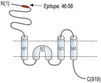
- Peptide (C)DGPNAQVMNAEEH, corresponding to amino acid residues 46-58 of rat kainate receptor GluK3 (Accession P42264). Extracellular, N-terminus.

 Western blot analysis of mouse (lanes 1 and 3) and rat (lanes 2 and 4) brain membranes:1,2. Anti-GRIK3 (GluK3) (extracellular) Antibody (#AGC-040), (1:200).
3,4. Anti-GRIK3 (GluK3) (extracellular) Antibody, preincubated with GRIK3/GluK3 (extracellular) Blocking Peptide (#BLP-GC040).
 Expression of GluR7 in mouse hippocampus and rat cerebellumImmunohistochemical staining of immersion-fixed, free floating frozen brain sections using Anti-GRIK3 (GluK3) (extracellular) Antibody (#AGC-040), (1:400). A. GluR7 staining (red) in mouse hippocampus reveals expression in the pyramidal (P) layer (arrow). B. GluR7 staining (red) in rat cerebellum reveals expression in the Purkinje (P) layer (horizontal arrow) and in the molecular layer (MOL) interneurons (arrow).
- Perrais, D. et al. (2009) Neuropharmacology 56,131
- Mayer, M.L. (2011) Curr. Opin. Neurobiol. 21, 283.
- Fernández-Alacid, L. et al. (2011) Eur. J. Neurosci. 34, 1724.
- Lerma, J. and Marques, J.M. (2013) Neuron 80, 292.
- Begni, S. et al. (2002) Mol. Psychiatry 7, 416.
- Schiffer, H.H. and Heinemann S.F. (2007) Am. J. Med. Genet. B. Neuropsychiatr. Genet. 144B, 20.
Ionotropic glutamate receptors (iGluRs) mediate the majority of fast excitatory synaptic neurotransmission in the central nervous system. The selective assembly of iGluRs into AMPA, kainate, and N-methyl-D-aspartic acid (NMDA) receptor subtypes is regulated by their extracellular amino-terminal domains (ATDs)1.
Kainate receptors (KARs) are tetramers classified into low-affinity receptor families (GluK1–GluK3) and high-affinity receptor families (GluK4–GluK5) based on their affinity for the neurotoxin kainic acid. KARs share a similar architecture with other ionotropic glutamate receptors; the subunits have a large extracellular domain composed of an amino-terminal domain (ATD) and a ligand binding domain (LBD) and an intracellular carboxy-terminal region2.
KARs are expressed in neurons and glial cells throughout the CNS. GluK3 is poorly expressed, appearing in layer IV of the neocortex and dentate gyrus in the hippocampus3.
KARs serve a crucial role as modulators of synaptic transmission and plasticity and their dysfunction has been linked to several disease states such as epilepsy, chronic pain and neurodegenerative diseases4. Alteration in the extracellular domain of GluK3, is in linkage disequilibrium with recurrent major depressive disorder patients5 and subjects with schizophrenia6.
Application key:
Species reactivity key:
Alomone Labs is pleased to offer a highly specific antibody directed against an extracellular epitope of the rat kainate receptor GluK3. Anti-GRIK3 (GluK3) (extracellular) Antibody (#AGC-040) can be used in western blot and immunohistochemistry applications. The antibody recognizes an extracellular epitope and is thus highly suited to recognize the receptor in living cells. It has been designed to recognize GluR7 from human, rat and mouse samples.
