Overview
- Peptide (C)STANRPNRPKSRESRDK, corresponding to amino acid residues 1184-1200 of mouse KCNMA1 (Accession number Q08460). Intracellular, C-terminal part.

 Western blot analysis of rat brain membranes:1. Anti-KCNMA1 (KCa1.1) (1184-1200) Antibody (#APC-107), (1:500).
2. Anti-KCNMA1 (KCa1.1) (1184-1200) Antibody, preincubated with KCNMA1/KCa1.1 (1184-1200) Blocking Peptide (#BLP-PC107).
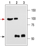
 Immunoprecipitation of rat brain lysate:1. Brain lysate.
2. Brain lysate immunoprecipitated with Anti-KCNMA1 (KCa1.1) (1184-1200) Antibody (#APC-107), (4 μg).
3. Brain lysate immunoprecipitated with pre-immune rabbit serum.
The upper arrow indicates the KCNMA1 channel while the lower arrow indicates the IgG heavy chain.
Western blot analysis was performed with Anti-KCNMA1 (KCa1.1) (1184-1200) Antibody.- Human cardiac fibroblasts (1:750) (Wang, Y.J. et al. (2006) J. Membrane Biol. 213, 175.).
 Expression of KCNMA1 in rat penisImmunohistochemical staining of rat penis transversal section using Anti-KCNMA1 (KCa1.1) (1184-1200) Antibody (#APC-107). Strong and specific immunostaining is evident in both corpus cavernosum smooth muscle cells (blue arrow) and in the muscular layer of the penis artery (green arrow). Universal Immuno-alkaline-phosphatase Polymer followed by New Fuchsin Subtrate (histofine, Nichirei Corp.) was used for the colour reaction. Hematoxillin is used as the counterstain.
- E9 chick CG neurons (Jha, S. and Dryer, S.T. (2009) FEBS Lett. 583, 3109.).
- Hofland L.J. and Lamberts, S.W.J. (2001) Ann. Oncol. 12, S31.
- Fombonne, J. et al. (2003) Rep. Biol. Endocrinol. 1, 19.
- Slooter, G.D. et al. (2001) Br. J. Surg. 88, 31.
- Schulz, S. et al. (2002) Gynecol. Oncol. 84, 235.
KCa1.1 (KCNMA1, BKCa, Maxi K+ or slo) is part of a structurally diverse group of K+ channels that are activated by an increase in intracellular Ca2+. KCa1.1 shows a large single channel conductance when recorded electrophysiologically and hence its name. It differs from the rest of the subfamily members in that it can be activated by both an increase in intracellular Ca2+ and by membrane depolarization.
KCa1.1 is expressed in virtually all cell types where it causes hyperpolarization and helps to connect intracellular Ca2+ signaling pathways and membrane excitability.
Indeed, KCa1.1 channels have a crucial role in smooth muscle contractility, neuronal spike shaping and neurotransmitter release.
Application key:
Species reactivity key:
Anti-KCNMA1 (KCa1.1) (1184-1200) Antibody (#APC-107) is a highly specific antibody directed against an epitope of the mouse protein. The antibody can be used in western blot, immunocytochemistry, immunohistochemistry, and immunoprecipitation applications. It has been designed to recognize KCNMA1 from human, rat, and mouse samples.
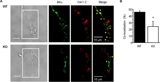
 Expression of BKCa in mouse mesenteric cells.A. Immunocytochemical staining of mouse mesenteric smooth muscle cells. Staining of BKCa using Anti-KCNMA1 (KCa1.1) (1184-1200) Antibody (#APC-107) (green) and CaV1.2 (red) in WT (upper) and Caveolin-1 KO (lower) mice. BKCa and CaV1.2 co-localization is shown in yellow (arrowheads). B. Ratio of BKCa and CaV1.2 co-localization particle number to total BKCa particle number in WT (n = 8) and KO (n = 7) myocytes (*, p < 0.05).Adapted from Suzuki, Y. et al. (2013) with permission of the American Society for Biochemistry and Molecular Biology.
Applications
Citations
- Western blot analysis and immunocytochemical staining of rat ROS17/2.8 osteoblasts. Tested in KCNMA1-/- cells.
Hei, H. et al. (2016) Mol. Cells 39, 530.
- Mouse podocyte lysate.
Wang, Y. et al. (2019) Front. Physiol. 10, 167. - Mouse bladder lysate.
Lu, M. et al. (2018) Am. J. Physiol. 314, C643. - Rat ROS17/2.8 osteoblast lysate. Also tested in KCNMA1-/- cells.
Hei, H. et al. (2016) Mol. Cells 39, 530. - Human dermal fibroblasts (1:200).
Kicinska, A. et al. (2016) Biochem. J. 473, 4457. - Mouse colon lysate.
Bhattarai, Y. et al. (2016) Am. J. Physiol. 311, G210. - Rat VSMC lysate (1:250).
Chen, M. et al. (2016) Physiol. Rep. 4, e12682. - Mouse kidney lysate.
Zhang, Y. et al. (2013) Am. J. Physiol. 305, F407. - Mitochondria and mitoplast from human endothelial EA.hy926 cells (1:200).
Bednarczyk, P. et al. (2013) Am. J. Physiol. 304, H1415.
- Rat VSMC.
Chen, M. et al. (2016) Physiol. Rep. 4, e12682. - Human CFBE cells.
Manzanares, D. et al. (2015) J. Biol. Chem. 290, 25710. - Human cardiac fibroblasts (1:750).
Wang, Y.J. et al. (2006) J. Membrane Biol. 213, 175.
- Mouse kidney sections (1:200).
Li, Y. et al. (2016) PLoS ONE 11, e0155006. - Human chorionic plate arterial smooth muscle cell sections (10 µg/ml).
Brereton, M. et al. (2013) PLoS ONE 8, e57451.
- Mouse podocytes.
Wang, Y. et al. (2019) Front. Physiol. 10, 167. - Rat ROS17/2.8 osteoblasts. Also tested in KCNMA1-/- cells.
Hei, H. et al. (2016) Mol. Cells 39, 530. - HEK 293 transfected cells (1:500).
Velazquez-Merrero, C. et al. (2016) J. Neurosci. 36, 10625. - Rat pinealocytes.
Mizutani, H. et al. (2016) Am. J. Physiol. 310, C740. - Rat hippocampal cells.
Palacio, S. et al. (2015) Alchol. Clin. Exp. Res. 39, 1619. - Human chorionic plate arterial smooth muscle cells (10 µg/ml).
Brereton, M. et al. (2013) PLoS ONE 8, e57451. - Mouse mesenteric smooth muscle cells (1:100).
Suzuki, Y. et al. (2013) J. Biol. Chem. 288, 36750. - E9 chick CG neurons.
Jha, S. and Dryer, S.T. (2009) FEBS Lett. 583, 3109.
- Wan, E. et al. (2013) FASEB J. 27, 1859.
- Yang, Y. et al. (2013) J. Physiol. 591, 1277.
- Howitt, L. et al. (2012) Am. J. Physiol. 302, H2464.
- Liu, Y. et al. (2012) Am. J. Physiol. 302, G44.
- Howitt, L. et al. (2011) Am. J. Physiol. 301, H29.
- Zuidema, M.Y. et al. (2010) Am. J. Physiol. 299, H1554.
- Grimes, W.N. et al. (2009) Nat. Neurosci. 12, 585.
- Li, Y. et al. (2009) BMC Dev. Biol. 9, 67.
- Yang, Y. et al. (2009) J. Physiol. 587, 3025.
- Rodriguez-Vilarrupla, A. et al. (2008) Liver Int. 28, 566.
- Ng, Y.K. et al. (2007) Am. J. Physiol. 292, R2100.
- Burnham, M. et al. (2006) Am. J. Physiol. 290, H1520.
