Overview
- Peptide (C)KLEE SLREQ AEKEG SALSV R, corresponding to amino acid residues 356-375 of rat Kir4.1 (Accession P49655). Intracellular, C-terminus.
- Rat brain membranes (1:400).
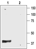
 Western blot analysis of rat brain membranes:1. Anti-Kir4.1 (KCNJ10) Antibody (#APC-035), (1:400).
2. Anti-Kir4.1 (KCNJ10) Antibody, preincubated with Kir4.1/KCNJ10 Blocking Peptide (#BLP-PC035).
- Rat renal cortex lysate (Cha, S.K. et al. (2011) J. Biol. Chem. 286, 1828.).
- Rat brain sections.
Mouse hippocampus (1:200) (Hsu, M.S. et al. (2011) Neuroscience 178, 21.).
Human brain tissue (Saadoun, S. et al. (2003) J. Clin. Pathol. 56, 972.).
- Mouse outer hair cells (OHCs) (Ruttiger, L. et al. (2004) Proc. Natl. Acad. Sci. U.S.A. 101, 12922.).
Kir4.1 is a member of the inward rectifying K+ channel family. The family includes 15 members that are structurally and functionally different from the voltage-dependent K+ channels.
The family’s topology consists of two transmembrane domains that flank a single and highly conserved pore region with intracellular N- and C-termini. As is the case for the voltage-dependent K+ channels the functional unit for the Kir channels is composed of four subunit that can assembly as either homo or heteromers.
Kir channels are characterized by a K+ efflux that is limited by depolarizing membrane potentials thus making them essential for controlling resting membrane potential and K+ homeostasis.
Kir4.1 is a member of the Kir4 subfamily that includes one other member: Kir4.2. Kir4.1 can co-assemble with Kir4.2 but also with other Kir channels such as Kir2.1 and Kir5.1.
The Kir4 subfamily has been classified as weak rectifiers with intermediate conductance.
Kir4.1, encoded by KCNJ10, is mainly expressed in brain, specifically in glia cells, but also in retina, ear and kidney.1,2
It has been proposed that Kir4.1 has an essential role in glial K+ buffering, a process that re-uptakes the K+ released during neuronal activity into the intracellular interstitial space. Loss of Kir4.1 causes retinal defects and loss of endochoclear potential.3
Application key:
Species reactivity key:

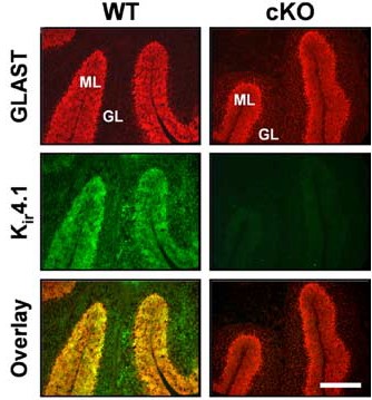
Knockout validation of Anti-Kir4.1 (KCNJ10) Antibody in mouse cerebellum.Immunohistochemical staining of mouse cerebellum sections using Anti-Kir4.1 (KCNJ10) Antibody (#APC-035). Kir4.1 staining (green). GLAST staining (red) co-localizes with Kir4.1 (overlay panel). Note the lack of Kir4.1 staining in Kir4.1 conditional knockout cerebellum.Adapted from Djukic, B. et al. (2007) J. Neurosci. 27, 11354. with permission of the Society for Neuroscience.
