Overview
- Peptide HDVLEVKRKYYKVNC, corresponding to amino acid residues 311-325 of rat KCNJ16 (Accession P52191). Intracellular, C-terminal.
- Rat kidney and liver membranes (1:200). Mouse brain and kidney membranes (1:200).
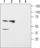
 Western blot analysis of rat kidney (lanes 1 and 3) and liver (lanes 2 and 4) membranes:1,2. Anti-Kir5.1 Antibody (#APC-123), (1:200).
3,4. Anti-Kir5.1 Antibody, preincubated with Kir5.1 Blocking Peptide (#BLP-PC123). Western blot analysis of mouse brain (lanes 1 and 3) and kidney (lanes 2 and 4) membranes:1,2. Anti-Kir5.1 Antibody (#APC-123), (1:200).
3,4. Anti-Kir5.1 Antibody, preincubated with Kir5.1 Blocking Peptide (#BLP-PC123).
- Rat kidney sections (paraffin embedded), (1:100).
Human brain sections (1:500) (Schirmer, L. et al. (2014) Ann. Neurol. 75, 810.).
Kir5.1 is a member of the family of inward rectifying K+ channels. The family includes 15 members that are structurally and functionally different from the voltage-dependent K+ channels.
The family’s protein topology consists of two transmembrane domains that flank a single and highly conserved pore region with intracellular N- and C-termini. As is the case for the voltage-dependent K+ channels the functional unit for the Kir channels is composed of four subunits that can assemble as either homo- or heteromers.
Kir channels are characterized by a K+ efflux that is limited by depolarizing membrane potentials thus making them essential for controlling resting membrane potential and K+ homeostasis.1
Kir5.1 can co-assemble with other Kir channels such as Kir2.1, Kir4.1 and Kir4.2. In fact, heteromeric Kir4.1/Kir5.1 form the basolateral small-conductance K+ channel in distal nephron segments that is responsible for generating the basolateral membrane potential that determines the magnitude and direction of K+ diffusion from cell to peritubular fluid in the nephron.2
The Kir5.1 homomeric channel was largely thought to be non-functional, although recent evidence demonstrates that Kir5.1 homomers can form functional channels when co-expressed with the anchor protein PSD-95 in the brain where it is abundantly expressed.3
Application key:
Species reactivity key:
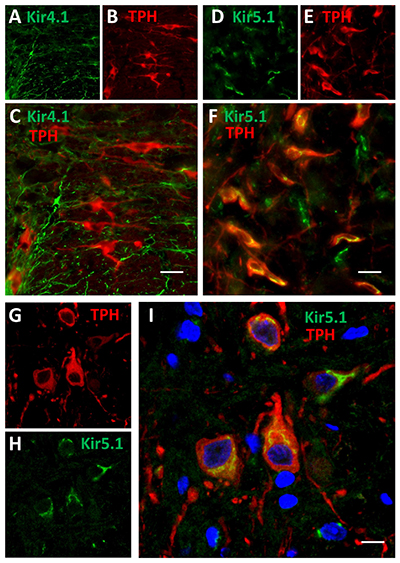
 Expression of Kir4.1 and Kir5.1 channels in rat brainstem.Immunohistochemical staining of rat brainstem sections using Anti-Kir4.1 (KCNJ10) Antibody (#APC-035) and Anti-Kir5.1 Antibody (#APC-123) showed that both channels are expressed (green). Kir4.1 staining is limited to astrocytes, while that of Kir5.1 is detected in 5-HT neurons together with the 5-HT neuronal marker TPH (red).Adapted from Puissant, M.M. et al. (2017) Front. Cell. Neurosci. 11, 34. with permission of Frontiers.