Overview
- Peptide (C)SVAVAKAKPKFSIS, corresponding to amino acid residues 372-385 of rat Kir6.2 (Accession P70673). Intracellular, C-terminal part.

 Western blot analysis of rat pancreas membranes:1. Anti-Kir6.2 Antibody (#APC-020), (1:200).
2. Anti-Kir6.2 Antibody, preincubated with Kir6.2 Blocking Peptide (#BLP-PC020).
- Mouse heart lysate (Li, J. et al. (2010) J. Biol. Chem. 285, 28723.).
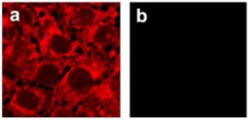
 Expression of Kir6.2 in rat pancreasImmunohistochemical staining of rat pancreas using Anti-Kir6.2 Antibody (#APC-020). A. Strong granular staining in a number of cells within the Islets of Langerhans is readily detected (red). B. The negative control slide shows no staining.
- Human placenta sections (Lybaert, P. et al. (2013) Placenta 34, 467.).
- Mouse ciliary cells (1:100) (Ohba, T. et al. (2013) J. Pharmacol. Exp. Ther. 347, 145.)
Mouse endothelial cells. Also tested in Kir6.2-/- mice (Milovanova, T. et al. (2005) Am. J. Physiol. 290, C66.).
- Babenko, A.P. et al. (1998) Annu. Rev. Physiol. 60, 667.
- Huopio, H. et al. (2002) Am. J. Physiol. 283, 207.
Kir6.2 is a member of the inward rectifier K+ channels (Kir channels), a large family of voltage-independent K+ channels largely involved in stabilization of the membrane resting potential and in K+ transport across membranes. Kir channels can be modulated by a variety of intracellular agents such as protons, GTP-binding proteins and adenine nucleotides.
The ATP-sensitive channel (KATP) is especially important since it couples cellular metabolism (intracellular ATP levels) with cell excitability. KATP channels have been described in pancreatic b-cells, neurons, heart, skeletal and smooth muscle.
The KATP channel is composed of a Kir6.2 or Kir6.1 subunit and a sulphonylurea receptor (SUR) subunit.
The pancreatic KATP channel for example, is composed of a complex of Kir6.2 and SUR1 subunits, while the cardiac KATP channel is composed of Kir6.2 and SUR2A complexes.
Impaired b-cell KATP channel function due to mutations in either Kir6.2 or SUR1 subunits has been linked to the recessive autosomal disorder called persistent hyperinsulinemic hypoglycemia of infancy (PHHI). In addition, a Kir6.2 variant has recently been linked to an increased risk of developing type-2 diabetes.
Application key:
Species reactivity key:
Anti-Kir6.2 Antibody (#APC-020) is a highly specific antibody directed against an epitope of the rat protein. The antibody can be used in western blot, immunocytochemistry, and immunohistochemistry applications. It has been designed to recognize Kir6.2 from human, rat, and mouse samples. The antibody is specific for Kir6.2 and does not cross react with Kir6.1.

Knockout validation of Anti-Kir6.2 Antibody in mouse endothelial cells.Immunocytochemical staining of mouse endothelial cells using Anti-Kir6.2 Antibody (#APC-020). A. Kir6.2 staining (red) in wild type cells. B. Kir6.2 immunoreactivity is not detected in Kir6.2-/- cells.Adapted from Milovanova, T. et al. (2005) Am. J. Physiol. 290, C66. with permission of The American Physiological Society.
Applications
Citations
 Expression of Kir6.2 in rat suprachiasmatic nucleus sectionsImmunohistochemical staining of rat suprachiasmatic nucleus sections (SCN) using Anti-Kir6.2 Antibody (#APC-020). Kir6.2 staining (green) colocalizes with Arginine-Vasopressin (AVP) staining (red) in the soma (middle panel) and in varicosities along the process (right panel).
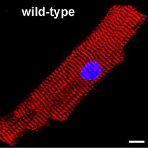
Adapted from Yang, J.J. et al. (2017) Sci. Rep. 7, 640. with permission of SPRINGER NATURE. Expression of Kir6.2 in mouse cardiomyocytes.Immunocytochemical staining of mouse cardiomyocytes using Anti-Kir6.2 Antibody (#APC-020).
Adapted from Li, J. et al. (2010) J. Biol. Chem. 285, 28723. with permission of The American Society for Biochemistry and Molecular Biology.
- Western blot analysis, immunocytochemistry, and indirect flow cytometry of mouse endothelial cells. Tested in Kir6.2-/- mice.
Milovanova, T. et al. (2005) Am. J. Physiol. 290, C66.
- Human myometrium tissue lysate.
Novakovic, R. et al. (2015) Mol. Hum. Reprod. 21, 545. - Rat immortalized chromaffin (MAH) cell lysate.
Salman, S. et al. (2014) Am. J. Physiol. 307, C266. - Human placenta and pancreas lysate.
Lybaert, P. et al. (2013) Placenta 34, 467. - Rat uterine tissue lysate.
Novakovic, R. et al. (2013) J. Physiol. Pharmacol. 64, 795. - Rat primary adrenomedullary chromafin cells (AMCs) (1:1000).
Salman, S. et al. (2013) J. Physiol. 591, 515. - Rat trigeminal ganglia lysate (1:500).
Niu, K. et al. (2011) Neuroscience 180, 344. - Rat heart lysate (1:200).
Yang, Z.W. et al. (2011) Acta Pharmacol. Sin. 32, 194. - Mouse heart lysate.
Li, J. et al. (2010) J. Biol. Chem. 285, 28723. - Rat DRG lysate (2010).
Zoga, V. et al. (2010) Mol. Pain 6, 1. - Mouse endothelial cell lysate. Also tested in Kir6.2-/- mice.
Milovanova, T. et al. (2005) Am. J. Physiol. 290, C66.
- Mouse heart lysate.
Li, J. et al. (2010) J. Biol. Chem. 285, 28723.
- Rat brain sections.
Yang, J.J. et al. (2017) Sci. Rep. 7, 640. - Human myometrium sections (1:50).
Novakovic, R. et al. (2015) Mol. Hum. Reprod. 21, 545. - Human placenta sections.
Lybaert, P. et al. (2013) Placenta 34, 467. - Human artery tissues (1:50).
Gojkovic-Bukarica, L. et al. (2011) Eur. J. Pharmacol. 654, 266. - Human HSV and HIMA tissues (1:50).
Gojkovic-Bukarica, L.C. et al. (2011) J. Cardiovasc. Pharmacol. 57, 648. - Rat trigeminal ganglia sections (1:1500).
Niu, K. et al. (2011) Neuroscience 180, 344. - Rat heart sections (1:100).
Yang, Z.W. et al. (2011) Acta Pharmacol. Sin. 32, 194. - Rat DRG sections (1:500).
Zoga, V. et al. (2010) Mol. Pain 6, 1.
- Mouse ciliary cells (1:100).
Ohba, T. et al. (2013) J. Pharmacol. Exp. Ther. 347, 145. - Mouse cardiomyocytes.
Li, J. et al. (2010) J. Biol. Chem. 285, 28723. - Mouse endothelial cells. Also tested in Kir6.2-/- mice.
Milovanova, T. et al. (2005) Am. J. Physiol. 290, C66.
- Mouse endothelial cells. Also tested in Kir6.2-/- mice.
Milovanova, T. et al. (2005) Am. J. Physiol. 290, C66.
- Xia, F. et al. (2004) J. Biol. Chem. 279, 24685.
