Overview
- Peptide (C)EMNGDLEIDHDVPPE, corresponding to amino acid residues 80-94 of rat KCNJ13 (Accession O70617). Extracellular loop.
- Rat kidney sections (paraffin-embedded), (1:50).
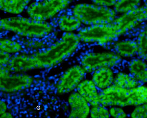
 Expression of Kir7.1 in rat kidneyImmunohistochemical staining of paraffin-embedded rat kidney sections using Anti-Kir7.1 (extracellular)-ATTO Fluor-488 Antibody (#APC-125-AG), (1:50). Kir7.1 staining (green) is present in convoluted tubules in the renal cortex. Glomeruli (G) are negative. Cell nuclei were visualized with Hoechst 33342 (blue).
Kir7.1 (KCNJ13) is a member of the inward rectifying K+ channel family. The family includes 15 members that are structurally and functionally different from the voltage-dependent K+ channels.
The family’s protein topology consists of two transmembrane domains that flank a single and highly conserved pore region with intracellular N- and C-termini. As is the case for the voltage-dependent K+ channels the functional unit for the Kir channels is composed of four subunits that can assemble as either homo- or heteromers.
Kir channels are characterized by a K+ efflux that is limited by depolarizing membrane potentials thus making them essential for controlling resting membrane potential and K+ homeostasis1.
Kir7.1, an inwardly rectifying K+ channel with unusual permeation properties is localized in epithelial cells of the thyroid, small intestine, kidney tubules, choroid plexus and in retinal pigment epithelium (RPE), where it forms a major component of the apical membrane K+ conductance2.
A mutation in the gene encoding the channel was found to cause snowflake vitreoretinal degeneration (SVD) which is a developmental and progressive hereditary eye disorder that affects multiple tissues within the eye3.
