Overview
- Peptide (C)EMNGDLEIDHDVPPE, corresponding to amino acid residues 80-94 of rat KCNJ13 (Accession O70617). Extracellular loop.

 Western blot analysis of rat brain membrane (lanes 1 and 4), mouse brain (lanes 2 and 5) and mouse kidney lysates (lanes 3 and 6):1-3. Anti-Kir7.1 (extracellular) Antibody (#APC-125), (1:200).
4-6. Anti-Kir7.1 (extracellular) Antibody, preincubated with Kir7.1 (extracellular) Blocking Peptide (#BLP-PC125).
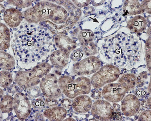
 Expression of Kir7.1 in rat kidneyImmunohistochemical staining of paraffin embedded section of rat kidney using Anti-Kir7.1 (extracellular) Antibody (#APC-125), (1:100). Staining is present in both distal (DT) and proximal (PT) tubules and in the collecting ducts (CD) in the renal cortex. Note that staining is absent both in glomeruli (Gl) and blood vessels (arrow). Hematoxilin is used as the counterstain.
- Bond, C.T. et al. (1994) Rec. Channels 2, 183.
- Yang, D. et al. (2008) Exp. Eye Res. 86, 81.
- Hejtmancik, J.F. et al. (2008) Am. J. Hum. Genet. 82, 174.
Kir7.1 (KCNJ13) is a member of the inward rectifying K+ channel family. The family includes 15 members that are structurally and functionally different from the voltage-dependent K+ channels.
The family’s protein topology consists of two transmembrane domains that flank a single and highly conserved pore region with intracellular N- and C-termini. As is the case for the voltage-dependent K+ channels, the functional unit for the Kir channels is composed of four subunits that can assemble as either homo- or heteromers.
Kir channels are characterized by a K+ efflux that is limited by depolarizing membrane potentials thus making them essential for controlling resting membrane potential and K+ homeostasis1.
Kir7.1, an inwardly rectifying K+ channel with unusual permeation properties is localized in epithelial cells of the thyroid, small intestine, kidney tubules, choroid plexus and in retinal pigment epithelium (RPE), where it forms a major component of the apical membrane K+ conductance2.
A mutation in the gene encoding the channel was found to cause snowflake vitreoretinal degeneration (SVD) which is a developmental and progressive hereditary eye disorder that affects multiple tissues within the eye3.
Application key:
Species reactivity key:
Anti-Kir7.1 (extracellular) Antibody (#APC-125) is a highly specific antibody directed against an epitope of the rat protein. The antibody can be used in western blot, immunohistochemistry and indirect flow cytometry applications. The antibody is directed against an extracellular epitope and is thus ideal for detecting Kir7.1 in living cells. It has been designed to recognize Kir7.1 from rat, mouse and human samples.

