Overview
- Peptide (C)RGPTITDKDRTKGPAE, corresponding to amino acid residues 578-593 of rat KCNQ2 (Accession O88943). Intracellular, C-terminus.
- KCNQ2 transfected HEK-293 cells (1:200). Human SHSY-5Y cells (1:1000) (Mucha, M. et al. (2010) J. Neurosci. 30, 13235.).
 Western blot analysis of KCNQ2 transfected HEK-293 cells:1. Anti-KCNQ2 Antibody (#APC-050), (1:200).
2. Anti-KCNQ2 Antibody, preincubated with KCNQ2 Blocking Peptide (#BLP-PC050).
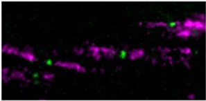
- Mouse optic nerve (Bekku, Y. et al. (2010) J. Neurosci. 30, 3113.).
- Rat cortical neurons (1:500) (Peretz, A. et al. (2005) Mol. Pharmacol. 67, 1053.).
The KCNQ family of voltage-gated K+ channels includes 5 known members: KCNQ1 to KCNQ5. Structurally, the KCNQ family belongs to the six transmembrane domain category of K+ channels.
KCNQ family members can form either homo- or heteromultimeric channels with different functional consequences. For example KCNQ2 and KCNQ3 heteromultimers give rise to a much larger channel current than when either protein is expressed alone, probably due to enhanced plasma membrane expression of the combined channel. Indeed, KCNQ2/KCNQ3 heteromultimers are believed to be the molecular correlates of the so-called M current. This current is a K+ neuronal current that is strongly inhibited by the activation of the M1 subtype of the muscarinic acetylcholine receptor.
Mutations in either KCNQ2 or KCNQ3 are associated with a form of epilepsy known as Benign familial neonatal convulsions (BNFC).
Application key:
Species reactivity key:
KCNQ2/Caspr
Expression of KCNQ2 (KV7.2) in mouse optic nerve.Immunohistochemical staining of mouse optic nerve sections using Anti-KCNQ2 Antibody (#APC-050). KCNQ2 staining (green) appears in the Nodes of Ranvier. Caspr staining is shown in purple.Adapted from Bekku, Y. et al. (2010) J. Neurosci. 30, 3113. with permission of the Society for Neuroscience.
