Overview
- Peptide AEGEKKEDNRYSDLKTIIC, corresponding to amino acid residues 668-686 of rat KCNQ3 (Accession O88944). Intracellular, C-terminal.

 Western blot analysis of rat brain membranes:1. Anti-KCNQ3 Antibody (#APC-051), (1:200).
2. Anti-KCNQ3 Antibody, preincubated with KCNQ3 Blocking Peptide (#BLP-PC051).
- Mouse cortical lysate (Ebner Bennatan, S. et al. (2012) J. Biol. Chem. 287, 27614.).
 Expression of KCNQ3 in rat hippocampusImmunohistochemical staining of perfusion-fixed frozen rat brain sections with Anti-KCNQ3 Antibody (#APC-051), (1:200), followed by goat anti-rabbit-AlexaFluor-488. KCNQ3 immunoreactivity (green) appears in cells of the pyramidal layer (P, vertical arrows) and cells of the stratum radiatum (SR, horizontal arrows). Cell nuclei are stained with DAPI (blue).
- Rat hippocampal neurons (1:100) (Rasmussen, H.B. et al. (2007) J. Cell Sci. 120, 953.).
- Kullmann, D.M. (2002) J. Mol. Brain 125, 1117.
- Robbins, J. (2001) Pharmacol. Ther. 90, 1.
- Wang, H.S. et al. (1998) Science 282, 1890.
The KCNQ family of voltage-gated K+ channels includes 5 known members: KCNQ1 to KCNQ5. Structurally, the KCNQ family belongs to the six transmembrane domain category of K+ channels. KCNQ family members can form either homomultimeric or heteromultimeric channels with different functional consequences. For example KCNQ2 and KCNQ3 heteromultimers give rise to a much larger channel current than when either protein is expressed alone, probably due to enhanced plasma membrane expression of the combined channel. Indeed, KCNQ2/KCNQ3 heteromultimers are believed to be the molecular correlates of the so-called M current. This current is a K+ neuronal current that is strongly inhibited by the activation of the M1 subtype of the muscarinic acetylcholine receptor.
Mutations in either KCNQ2 or KCNQ3 are associated with a form of epilepsy known as benign familial neonatal convulsions (BFNC).
Application key:
Species reactivity key:
Anti-KCNQ3 Antibody (#APC-051) is a highly specific antibody directed against an epitope of the rat KV7.3 channel. The antibody can be used in western blot, immunoprecipitation, immunohistochemistry, and immunocytochemistry applications. It has been designed to recognize KCNQ3 from rat, human, and mouse samples.

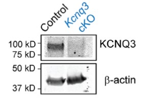
Knockout validation of Anti-KCNQ3 Antibody in mouse hippocampus.Western blot analysis of mouse hippocampus membrane fractions using Anti-KNQ3 Antibody (#APC-051), (upper panel). KCNQ3 immunodetection is observe in control mice (left lane) and absent in KCNQ3 conditional knockout mice (right lane). β-actin is used as a loading control.Adapted from Soh, H. et al. (2014) J. Neurosci. 34, 5311. with permission of the Society for Neuroscience.
Applications
Citations
 Expression of KCNQ3 in guinea pig bladder detrusor smooth muscleImmunohistochemical staining of guinea pig bladder detrusor smooth muscle sections using Anti-KCNQ3 Antibody (#APC-051). KCNQ3 staining (green) is detected in smooth muscle bundles (SM) and in interstitial cells (IC) adjacent to and between SM. Inset: pre-absorption control.
Adapted from Anderson, U.A. et al. (2013) Br. J. Pharmacol. 169, 1290. with permission of John Wiley & Sons.
- Western blot analysis of mouse hippocampus lysate (1:200). Tested in conditional knockout KCNQ3 brain lysate.
Soh, H. et al. (2014) J. Neurosci. 34, 5311.
- Rat brain lysate.
Parrilla-Carrero, J. et al. (2018) J. Neurosci. 38, 4212. - CHO transfected cells.
Ambrosino, P. et al. (2018) Mol. Neurobiol. 55, 7009. - Mouse brain lysate.
Paz, R.M. et al. (2018) Neuropharmacology 137, 309. - HEK 293 transfected cells.
Yau, M.C. et al. (2018) J. Gen. Physiol. 150, 1421. - Rat brain PVN membrane fraction(1:1000).
Zhou, J.J. et al. (2017) Neuropharmacology 114, 67. - CHO transfected cells (1:1000).
Miceli, F. et al. (2015) J. Neurosci. 35, 3782. - Mouse hippocampus lysate (1:200). Also tested in conditional knockout KCNQ3 brain lysate.
Soh, H. et al. (2014) J. Neurosci. 34, 5311.
- Mouse cortical lysate.
Ebner Bennatan, S. et al. (2012) J. Biol. Chem. 287, 27614.
- Mouse brain sections.
Tirko, N.N. et al. (2018) Neuron 100, 593. - Mouse brain sections (1:100).
Paz, R.M. et al. (2018) Neuropharmacology 137, 309. - Rat brainstem sections (1:100).
Ghezzi, F. et al. (2018) J. Physiol. 596, 2611. - Rat brain sections.
Zhou, J.J. et al. (2017) Neuropharmacology 114, 67. - Guinea pig detrusor (1:200).
Anderson, U.A. et al. (2013) Br. J. Pharmacol. 169, 1290.
- Rat hippocampal neurons (1:100).
Rasmussen, H.B. et al. (2007) J. Cell Sci. 120, 953.
- Bréchet, A. et al. (2008) J. Cell Biol. 183, 1101.
- Kanaumi, T. et al. (2008) Brain Devel. 30, 362.
- Wladyka, C.L. et al. (2008) J. Physiol. 586, 795.
- Peretz, A. et al. (2007) J. Neurophysiol. 97, 283.
- Peretz, A. et al. (2005) Mol. Pharmacol. 67, 1053.
- Jiang, M. et al. (2004) Circulation 109, 1783.
