Overview
- Peptide (C)DRKTREKGDKGPSD, corresponding to amino acid residues 594-607 of mouse KCNQ4 (Accession Q9JK97). Intracellular, C-terminus.
- Rat and mouse brain membranes (1:200-1:1000).
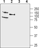
 Western blot analysis of rat (lanes 1 and 3) and mouse (lanes 2 and 4) brain membranes:1,2. Anti-KCNQ4 Antibody (#APC-164), (1:200).
3,4. Anti-KCNQ4 Antibody, preincubated with KCNQ4 Blocking Peptide (#BLP-PC164).
- Rat brain sections (1:400).
The KCNQ (KV7) family of channels consists of five members (KCNQ1, KCNQ2, KCNQ3, KCNQ4 and KCNQ5). KCNQ channels commonly display activation at voltages close to neuronal resting membrane potentials and are regulated by G-protein coupled receptor (GPCR) signaling, notably by muscarinic receptors1.
The KCNQ4 (KV7.4) encodes a protein of 695 amino acid residues. The homology with other members of the KCNQ family ranges from 37 to 44% at the amino acid level. KCNQ4 protein contains six domains that span the plasma membrane, and a P-loop domain which forms the K+-selective channel pore. The fourth transmembrane domain contains the voltage sensor, which is responsible for the electrically driven conformational change that leads to channel opening. Functional channels are formed by a tetrameric assembly of KCNQ4 subunits, typically in homotetrameric form, but heterotetrameric co-assembly of different members is allowed2.
KCNQ2-KCNQ5 are predominantly expressed in the peripheral and central nervous system, including hippocampal cells, cortical cells, and dorsal root ganglion3. In addition KCNQ4 is expressed in the heart and in the outer, but not in the inner, sensory hair cells of the cochlea4.
It has been demonstrated that KCNQ4 mutations may be a relatively frequent cause of autosomal dominant hearing loss4.
