Overview
- Peptide (C)RGSQDFYPKWRESK, corresponding to amino acid residues 856-869 of mouse KCNQ5 (Accession Q9JK45). Intracellular, C-terminus.

 Western blot analysis of rat hippocampus (lanes 1 and 4), mouse brain (lanes 2 and 5) and rat brain (lanes 3 and 6) lysates:1-3. Anti-KCNQ5 Antibody (#APC-155), (1:200).
4-5. Anti-KCNQ5 Antibody, preincubated with KCNQ5 Blocking Peptide (#BLP-PC155).
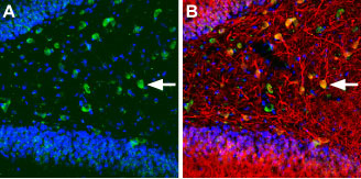
 Expression of KCNQ5 in rat hippocampusImmunohistochemical staining of immersion-fixed, free floating rat brain frozen sections using Anti-KCNQ5 Antibody (#APC-155), (1:100). A. KCNQ5 (green) is expressed in hilar cells of the hippocampal dentate gyrus. B. Double-staining of KCNQ5 (green) with a neuronal marker (“Milli-Mark” from Millipore, red) reveals partial localization with hilar interneurons (arrow points at an example).
- Rat smooth muscle cells (1:100) (Morales-Cano, D. et al. (2015) Cardiovasc. Res. 106, 98.).
- Brown, D.A. and Passmore, G.M. (2009) Br. J. Pharmacol. 156, 1185.
- Jentsch, T.J. (2000) Nat. Rev. Neurosci. 1, 21.
- Robbins, J. (2001) Pharmacol. Ther. 90, 1.
- Jensen, H.S. et al. (2005) Brain Res. Mol. Brain Res. 139, 52.
- Lerche, C. et al. (2000) J. Biol. Chem. 275, 22395.
KCNQ genes encode members of the KV7 K+ channels. Structurally, the KCNQ family belongs to the six transmembrane domain category of K+ channels. Five members belong to this family (KCNQ1-KCNQ5) of which KCNQ2 to KCNQ5 are expressed in the nervous system1-3. KCNQ5 is also expressed in visceral smooth muscle4 and skeletal muscle5.
These channels form subunits of the low threshold voltage gated K+ channel. KCNQ2 and KCNQ3 mostly form heteromers to form the M current. KCNQ5 is known to contribute to this current1.
Application key:
Species reactivity key:
Anti-KCNQ5 Antibody (#APC-155) is a highly specific antibody directed against an epitope of the mouse KV7.5 channel. The antibody can be used in western blot, immunohistochemistry, and immunocytochemistry applications. It has been designed to recognize KCNQ5 from rat, mouse, and human samples.

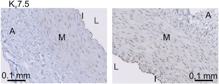
Expression of KV7.5 (KCNQ5) in swine coronary artery.Immunohistochemical staining of swine coronary artery sections using Anti-KCNQ5 Antibody (#APC-155). KV7.5 staining (brown) is mostly detected in the intimal layer (I). Much lower staining is observed in medial layer (M).Adapted from Chen, X. et al. (2016) PLoS ONE 11, e0148569. with permission of PLoS.
Applications
Citations
 Expression of KCNQ1 (KV7.1) and KCNQ5 (KV7.5) in rat smooth muscle cells.Immunocytochemical staining of rat smooth muscle cells from LCA and RCA using Anti-KCNQ1 Antibody (#APC-022) and Anti-KCNQ5 Antibody (#APC-155) shows that the expression of both channels is higher in LCA than in RCA.
Adapted from Morales-Cano, D. et al. (2015) with permission of the European Society of Cardiology.
- Rat brain lysate.
Parrilla-Carrero, J. et al. (2018) J. Neurosci. 38, 4212. - Mouse brain lysate.
Paz, R.M. et al. (2018) Neuropharmacology 137, 309.
- Rat brainstem sections (1:400).
Ghezzi, F. et al. (2018) J. Physiol. 596, 2611. - Mouse brain sections (1:100).
Paz, R.M. et al. (2018) Neuropharmacology 137, 309. - Swine coronary artery sections.
Chen, X. et al. (2016) PLoS ONE 11, e0148569.
- Rat smooth muscle cells (1:500).
Morales-Cano, D. et al. (2015) Cardiovasc. Res. 106, 98.
