Overview
- Peptide CKGEFFWLEPKNAFEN, corresponding to amino acid residues 211 - 226 of rat SLC7A8 (Accession Q9WVR6). Extracellular, 3rd loop.

 Western blot analysis of mouse brain lysates (lanes 1 and 3) and rat brain synaptosomal fraction (lanes 2 and 4):1-2. Anti-LAT2 (SLC7A8)(extracellular) Antibody (#ANT-108), (1:200).
3-4. Anti-LAT2 (SLC7A8)(extracellular) Antibody, preincubated with LAT2 (SLC7A8)(extracellular) Blocking Peptide (BLP-NT108). Western blot analysis of human Jurkat T-cell leukemia cell line lysates (lanes 1 and 4), human MDA-MB-231 breast adenocarcinoma cell line lysates (lanes 2 and 5) and human MCF-7 breast adenocarcinoma cell line lysates (lanes 3 and 6):1-3. Anti-LAT2 (SLC7A8)(extracellular) Antibody (#ANT-108), (1:200).
4-6. Anti-LAT2 (SLC7A8)(extracellular) Antibody, preincubated with LAT2 (SLC7A8)(extracellular) Blocking Peptide (BLP-NT108).
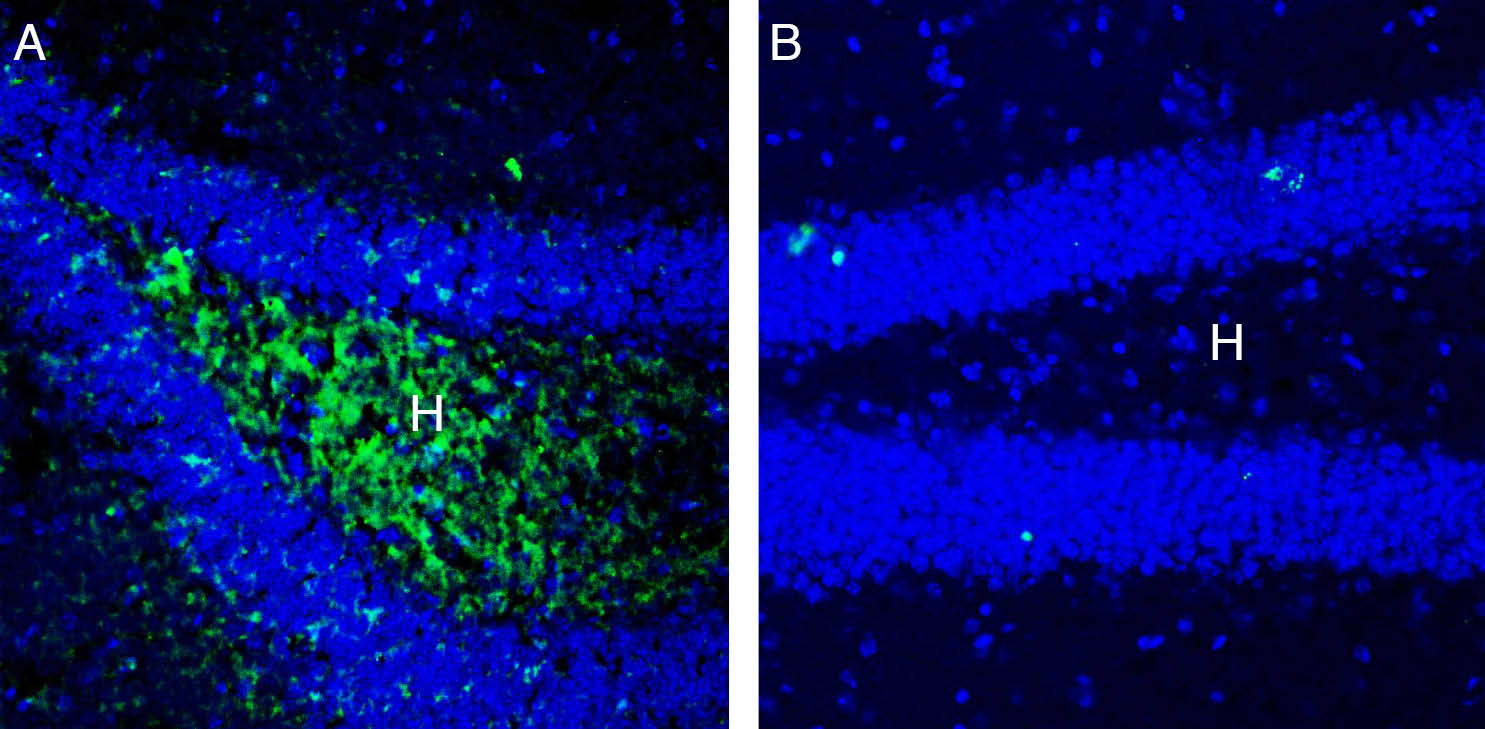
 Expression of SLC7A8 in mouse hippocampus.Immunohistochemical staining of perfusion-fixed frozen rat brain sections with Anti-LAT2 (SLC7A8)(extracellular) Antibody (#ANT-108), (1:200), followed by goat anti-rabbit-AlexaFluor-488. A. Staining in the mouse hippocampal dentate gyrus region, showed immunoreactivity of SLC7A8 (green) in the hilus (H). B. Pre-incubation of the antibody with LAT2 (SLC7A8)(extracellular) Blocking Peptide (BLP-NT108), suppressed staining. Cell nuclei are stained with DAPI (blue).
- Wang et al (2015) Am J Cancer Res, 15, 1281.
- Krause et al (2017) Mol Cell Endocrinol 15, 68.
- Del Amo et al (2008) J Pharmacol Sci 119, 368.
- Espino Guarch et al (2018) eLife 7, e31511.
- Knöpfel EB et al (2019) Front. Physiol. 10, 688.
L-type amino acid transporter (LAT) family are transporters responsible for the uptake of neutral amino acids into cells. The LATs family contain four different members LAT1 (SLC7A5), LAT2 (SLC7A8), LAT3 (SLC43A1) and LAT4 (SLC43A2). LATs transporters are known to carried out their function in an Na+ and pH independent manner1. In recent years, LATs family shown to participate in the uptake of thyroid hormones (THs) and their derivatives2.
LAT2 (SLC7A8) is a transmembrane protein first discovered back in 1999 using sequence similarity to LAT1. According to the predicted membrane topology, LAT2 consists of 12 transmembrane domains (TMDs) and N- and C- termini located in the cytosol. LAT2 associate with the 4F2hc (4F2 antigen heavy chain; CD98 heavy chain) glycoprotein to form a dimer that act as a neutral amino acid transporter3.
LAT2 is expressed in various tissues, including the intestinal wall, blood–brain barrier, and kidney. Mutations in LAT2 protein cause age-related hearing loss in mice and humans5. In addition, deletion LAT2 in mice led to increased incidence of cataract4,5.
Application key:
Species reactivity key:
Anti-LAT2 (SLC7A8) (extracellular) Antibody (#ANT-108) is a highly specific antibody directed against an extracellular epitope of the rat protein. The antibody can be used in western blot, immunohistochemistry and flow cytometry applications. It has been designed to recognize SLC7A8 from rat, mouse and human samples.

