Overview
- Peptide (C)HVRSYSPDWPHQPNK, corresponding to amino acid residues 507-521 of mouse LINGO-1 (Accession Q9D1T0). Extracellular, N-terminus.

 Western blot analysis of rat (lanes 1 and 3) and mouse (lanes 2 and 4) brain membranes:1,2. Anti-LINGO-1 (extracellular) Antibody (#ANT-032), (1:200).
3,4. Anti-LINGO-1 (extracellular) Antibody, preincubated with LINGO-1 (extracellular) Blocking Peptide (#BLP-NT032). Western blot analysis of human SH-SY5Y neuroblastoma cell line lysate:1. Anti-LINGO-1 (extracellular) Antibody (#ANT-032), (1:500).
2. Anti-LINGO-1 (extracellular) Antibody, preincubated with LINGO-1 (extracellular) Blocking Peptide (#BLP-NT032).
 Expression of LINGO-1 in rat brain and spinal cordImmunohistochemical staining of perfusion-fixed frozen rat brain sections using Anti-LINGO-1 (extracellular) Antibody (#ANT-032), (1:400), followed by donkey-anti-rabbit-Cy3. A. LINGO-1 staining (red) is detected in sections of rat substantia nigra, in nigral neurons (arrows). B. Staining of rat spinal cord shows LINGO-1 labeling in ventral horn neurons (arrows). Cell nuclei are stained with DAPI (blue).
LINGO-1 (LRR and Ig domain-containing, nogo receptor interacting protein) is a protein highly expressed in the brain but not in other tissues. There are currently two other known human paralogs- LINGO-2 and LINGO-3. LINGO-1 contains 12 leucine-rich repeat (LRR) motifs flanked by N- and C- terminal capping domains, one immunoglobulin (Ig) domain, a transmembrane domain and a short cytoplasmic tail. The cytoplasmic tail contains a canonical epidermal growth factor receptor-like tyrosine phosphorylation site. LINGO-1 is highly conserved evolutionary with human and mouse orthologues, sharing 99.5% identity. LINGO-1 binds both to Nogo-66 receptor (NgR1) and to p75 neurotrophin receptor simultaneously and can form a ternary complex1.
In animal models, LINGO-1 expression is upregulated in rat spinal cord injury, experimental autoimmune encephalomyelitis, neurotoxic lesions and glaucoma. In humans, LINGO-1 expression is increased in oligodendrocyte progenitor cells from demyelinated white matter of multiple sclerosis post-mortem samples, and in dopaminergic neurons from Parkinson’s disease brains. LINGO-1 negatively regulates oligodendrocyte differentiation and myelination, neuronal survival and axonal regeneration by activating the RhoA and inhibiting protein kinase B (Akt) phosphorylation signaling pathways2.
Application key:
Species reactivity key:
Alomone Labs is pleased to offer a highly specific antibody directed against an extracellular epitope of mouse LINGO-1. Anti-LINGO-1 (extracellular) Antibody (#ANT-032) can be used in western blot and immunohistochemistry applications. The antibody recognizes an extracellular epitope, and is thus ideal for detecting the protein in living cells. It has been designed to recognize LINGO-1 from human, mouse, and rat samples.

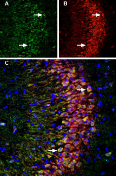
Multiplex staining of LINGO-1 and p75NTR in rat hippocampus.Immunohistochemical staining of immersion-fixed, free floating rat brain frozen sections using Anti-LINGO-1 (extracellular) Antibody (#ANT-032), (1:200) and Anti-p75 NGF Receptor (extracellular)-ATTO Fluor-550 Antibody (#ANT-007-AO), (1:80). A. LINGO-1 staining (green) in hippocampal CA3 region is detected in neuronal profiles (arrows). B. p75NTR staining in the same section (orange) is also seen in CA3 neurons (arrows). C. Merge of the two images demonstrates colocalization in several neurons (arrows). Cell nuclei are stained with DAPI (blue).
