Overview
- Peptide (C)HAQGIARLHKRQRPVH, corresponding to amino acid residues 217-232 of human MC1R (Accession Q01726). 3rd intracellular loop.

 Western blot analysis of human normal skin fibroblast cell line Malme-3 (lanes 1 and 4) and human malignant melanoma cell lines Malme-3M (lanes 2 and 5) and A875 (lanes 3 and 6):1,2,3. Anti-MC1 Receptor Antibody (#AMR-020), (1:500).
4,5,6. Anti-MC1 Receptor Antibody, preincubated with MC1 Receptor Blocking Peptide (#BLP-MR020). Western blot analysis of rat adrenal lysate:1. Anti-MC1 Receptor Antibody (#AMR-020), (1:400).
2. Anti-MC1 Receptor Antibody, preincubated with MC1 Receptor Blocking Peptide (#BLP-MR020).
 Expression of MC1R in normal skin and melanomaImmunohistochemical staining of paraffin embedded normal skin and melanoma sections using Anti-MC1 Receptor Antibody (#AMR-020) (1:100). MCR1 staining (red-brown color is highly specific in A. epidermal cells, B. eccrine sweat gland cells and C. melanoma cells. Color reaction was obtained with DAB. Hematoxilin is used as the counterstain.
 Expression of MC1R in human Malme-3M cellsImmunocytochemical staining of human paraformaldehyde fixed and permeabilized malignant melanoma cell lines (Malme-3M). A. Cells were stained with Anti-MC1 Receptor Antibody (#AMR-020), (1:200) followed by goat-anti-rabbit-AlexaFluor-555 secondary antibody. B. Live view of the same field as in (A). C. Computer merge of (A) and (B).
- Abdel-Malek, Z.A. (2001) Cell. Mol. Life Sci. 58, 434.
- Gantz, I. and Fong, T.M. (2003) Am. J. Physiol. 284, E468.
- Wikberg, J.E. et al. (2000) Pharm. Res. 42, 393.
- Dinulescu, D.M. and Cone, R.D. (2000) J. Biol. Chem. 275, 6695.
- Kanetsky, P.A. et al. (2006) Cancer Res. 66, 9330.
- Catania, A. et al. (2004) Pharmacol. Rev. 56, 1.
Melanocortin receptor 1 (MC1R) belongs to a five-member receptor family known as the melanocortin receptors. The melanocortin receptors are members of the 7-transmembrane domain, G protein-coupled receptor (GPCR) superfamily.
The receptors ligands, the melanocortins, are a group of structurally derived peptides consisting of α-, β- and γ-melanocyte stimulating hormone (α, β, γ-MSH) and the adrenocorticotropic hormone (ACTH) all of which are derived from the post-translational processing of a common precursor peptide, proopiomelanocortin (POMC).1,2,3
One of the most salient features of the melanocortin signaling system is the presence of two endogenous antagonists, that is proteins that bind specifically to the receptor but instead of activating it have an inhibitory effect. The antagonist proteins are termed agouti (or agouti signaling protein, ASP) and agouti-related protein (AGRP).4
All five melanocortin receptors bind their agonists (the melanocortins) and their endogenous antagonists (agouti and AGRP) with different affinities.
MC1R was the first member of the melanocortin receptor family to be cloned. The receptor transduces signals via Gs resulting in the activation of adenylate cyclase and production of cAMP.
MC1R binds its ligands with the following potency: α-MSH = ACTH > β-MSH > γ-MSH. MC1R also binds the endogenous antagonist agouti with high affinity.1, 2
MC1R can be described as the “classical” melanocyte α-MSH receptor. The receptor is expressed in the skin where it has a key role in determining skin and hair pigmentation. In fur-bearing mammals, the local ratio between α-MSH and agouti will determine coat pigmentation as α-MSH stimulates and agouti inhibits melanin production.
In humans, especially in Caucasians, the MC1R gene is highly polymorphic and several allelic variants have been correlated with red-hair, poor tanning ability and increased risk of melanoma.1,3,5 In addition, MC1R expression in melanoma has been shown to be upregulated up to 20-fold when compared to normal melanocytes.
The melanocortins and particularly α-MSH have significant anti-inflammatory properties. Since α-MSH binds to MC1R with the highest potency, it was proposed that the latter mediated the anti-inflammatory effects of α-MSH. Indeed, MC1R expression has been demonstrated in several cells of the immune system including macrophages and neutrophils.6
Application key:
Species reactivity key:
Alomone Labs is pleased to offer a highly specific antibody directed against an epitope of the human melanocortin receptor 1. Anti-MC1 Receptor Antibody (#AMR-020) can be used in western blot, immunocytochemistry and immunohistochemistry applications. It has been designed to recognize MC1R from human and rat samples. The antibody is unlikely to recognize MC1R from mouse samples.

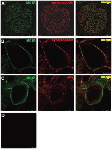
Expression of MC1R in human kidney.Immunohistochemical staining of human kidney biopsy sections using Anti-MC1 Receptor Antibody (#AMR-020). A. MC1R staining (green) is detected in the glomerular and immune-colocalizes with synaptopodin, a podocyte marker. B. Enlarged images of glomerular capillaries. C. MC1R staining does not colocalize with endothelial-specific lectin Ulex europaeus agglutinin I, an endothelial cell marker. D. Primary antibody was omitted.Adapted from Lindskog, A. et al. (2010) J. Am. Soc. Nephrol. 21, 1290. with permission of the American Society of Nephrology.
Applications
Citations
- Western blot of rat kidney. Tested in rats with Crispr/Cas9 generated MC1R knockouts.
Chen, B. et al. (2023) J. Am. Soc. Nephrol. 34, 467.
- Rat liver lysate (1:500).
Lonati, C. et al. (2014) Peptides 50, 145. - Rat kidney and testis.
Si, J. et al. (2013) Kidney Int. 83, 635. - Human kidney lysate (1:500).
Lindskog, A. et al. (2010) J. Am. Soc. Nephrol. 21, 1290.
- Rat kidney sections.
Si, J. et al. (2013) Kidney Int. 83, 635. - Human kidney sections (1:100).
Lindskog, A. et al. (2010) J. Am. Soc. Nephrol. 21, 1290.
- Rat renal tubular epithelial cells.
Si, J. et al. (2013) Kidney Int. 83, 635.
- Shen, Y. et al. (2013) J. Neurosci. 33, 464.
