Overview
- Peptide (C)HRGMHTSLHLWNRSS, corresponding to amino acid residues 6-20 of human MC4R (Accession P32245). Extracellular, N terminal.

 Western blot analysis of rat brain lysates:1. Anti-MC4 Receptor (extracellular) Antibody (#AMR-024), (1:400).
2. Anti-MC4 Receptor (extracellular) Antibody, preincubated with MC4 Receptor (extracellular) Blocking Peptide (#BLP-MR024). Western blot analysis of mouse brain lysates:1. Anti-MC4 Receptor (extracellular) Antibody (#AMR-024), (1:400).
2. Anti-MC4 Receptor (extracellular) Antibody, preincubated with MC4 Receptor (extracellular) Blocking Peptide (#BLP-MR024).
 Expression of MC4R in mouse brainImmunohistochemical staining of perfusion-fixed frozen mouse brain sections using Anti-MC4 Receptor (extracellular) Antibody (#AMR-024), (1:100). MC4R (green) is expressed in the mouse hypothalamus in axonal processes (arrows). Hoechst 33342 is used as the counterstain (blue).
 Expression of MC4R in rat pituitary cell lineCell surface detection of MC4R in live intact GH3 cells with Anti-MC4 Receptor (extracellular) Antibody (#AMR-024), (1:50). A. MC4R staining was observed, followed by Alexa-555-conjugated goat anti-rabbit secondary antibody (red). Hoechst 33342 (blue) is used to visualize the nuclei. B. Live view of the same field as A.
- Abdel-Malek, Z.A. (2001) Cell. Mol. Life Sci. 58, 434.
- Gantz, I. and Fong, T.M. (2003) Am. J. Physiol. 284, E468.
- Wikberg, J.E. et al. (2000) Pharmacol. Res. 42, 393.
- Huszar, D. et al (1997) Cell 88, 131.
- Adan, R.A. et al. (2006) Br. J. Pharmacol. 149, 815.
Melanocortin receptor 4 (MC4R) is one of five members of the melanocortin receptor family, which belongs to the 7-transmembrane domain, G protein-coupled receptor (GPCR) superfamily.
The ligands of these receptors, the melanocortins, are a group of structurally-related peptides comprising the α-, β-, and γ-melanocyte-stimulating hormone (α-, β-, γ-MSH) and the adrenocorticotropic hormone (ACTH), all of which are derived from post-translational processing of a common precursor peptide, proopiomelanocortin (POMC).1,2,3
One of the salient features of the melanocortin signaling system is the existence of two endogenous antagonists, proteins that bind specifically to the receptor and have an inhibitory effect. These antagonist proteins are termed agouti (or agouti signaling protein, ASP) and agouti-related protein (AGRP).1
All five melanocortin receptors bind their agonists (the melanocortins) and their endogenous antagonists (agouti/ASP and AGRP) with differing affinities. The order of potency for MC4R activation is α-MSH = ACTH > β-MSH >> γ-MSH. AGRP and agouti/ASP are both endogenous antagonists of MC4R.1,2,3
MC4R is widely expressed in the brain including the cortex, thalamus, hypothalamus, and brain stem. The best understood physiological role of MC4R is in the regulation of feeding behavior and the control of body weight. Indeed, mice with a targeted disruption of the MC4R gene display an obese phenotype characterized by increased adiposity, hyperphagia, and hyperleptinaemia.4 Similarly, MC4R mutations in humans have been associated with severe childhood obesity including characteristics very similar to the MC4R knockout mouse phenotype.5 All these make MC4R an attractive drug target for the treatment of obesity.
Application key:
Species reactivity key:
Anti-MC4 Receptor (extracellular) Antibody (#AMR-024) is a highly specific antibody directed against an epitope of the human melanocortin receptor 4. The antibody can be used in western blot, immunohistochemistry, and immunocytochemistry applications. It recognizes an extracellular epitope and can potentially recognize the receptor in living cells. It has been designed to recognize MC4R from human, mouse, and rat samples.

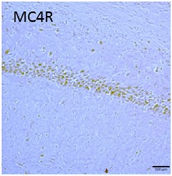
Expression of MC4R in rat hippocampus. Immunohistochemical staining of rat brain sections using Anti-MC4 Receptor (extracellular) Antibody (#AMR-024). MC4R staining (brown) is detected in CA1 region. Adapted from Massey, A.T. et al. (2016) Front. Neurol. 7, 65. with permission of Frontiers.
Applications
Citations
 Expression of MC4R in mouse hippocampal neurons.A. Immunocytochemical staining of mouse hippocampal neurons. Extracellular staining of cells using Anti-MC4 Receptor (extracellular) Antibody (#AMR-024) (green). B. Western blot analysis of MC4R-transfected HEK 293 cells treated with shRNA targeting MC4R. shRNA treatment decreased the receptor level by 83%. MC4R protein levels were detected with Anti-MC4 Receptor (extracellular) Antibody.
Adapted from Shen, Y. et al. (2013) with permission of the Society for Neuroscience.
- Rat brain sections.
Massey, A.T. et al. (2016) Front. Neurol. 7, 65.
- Mouse hippocampal neurons.
Shen, Y. et al. (2013) J. Neurosci. 33, 464.
- Peng, C.Y. et al. (2012) J. Neurosci. 32, 17211.
