Overview
- Peptide (C)EFERHDPVDGRISER, corresponding to amino acid residues 313-327 of rat MICU1 (Accession Q6P6Q9). Intracellular.
- Rat skeletal muscle and brain lysates. Mouse kidney lysates (1:200-1:1000).
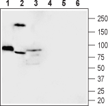
 Western blot analysis of rat skeletal muscle (lanes 1 and 4), mouse kidney (lanes 2 and 5) and rat brain (lanes 3 and 6) lysates:1-3. Anti-MICU1 Antibody (#ACC-322), (1:200).
4-6. Anti-MICU1 Antibody, preincubated with MICU1 Blocking Peptide (#BLP-CC322).
Mitochondrial Ca2+ uptake was found to be mediated by the mitochondrial Ca2+ uniporter (MCU). MCU is a ruthenium‐red‐sensitive and highly selective Ca2+ ion channel that uptakes massive amount of Ca2+ across the inner membrane1. MICU1 is a ~54 kDa. protein with an amino‐terminal mitochondrial targeting sequence, a predicted transmembrane helix, and a cytosolic C‐terminus containing two classical EF‐hand Ca2+‐binding domains2.
Mitochondrial calcium uptake 1 (MICU1) protein was identified as an essential element of mitochondrial Ca2+ uptake. MCU initially takes up Ca2+ very rapidly, but when [Ca2+] inevitably increases inside the matrix, MICU1 binds Ca2+ through its EF‐hand domains, exerting an inhibitory role on MCU‐dependent Ca2+ entry3.
RNA expression of MICU1 is strongly correlated across a variety of tissues including: stomach, kidney, small and large intestine, cerebral cortex, cerebellum, hypothalamus, spinal cord and skeletal muscles4.
Loss-of-function mutations in MICU1 causes a brain and muscle disorder linked to primary alterations in mitochondrial Ca2+ signaling5.
