Overview
- Peptide (C)GRLNDMYGDYKYT, corresponding to amino acid residues 403-415 of rat MCT1 (Accession P53987). 6th extracellular loop.

 Western blot analysis of rat (lanes 1 and 3) and mouse (lanes 2 and 4) brain lysates:1,2. Anti-MCT1 (SLC16A1) (extracellular) Antibody (#AMT-011), (1:200).
3,4. Anti-MCT1 (SLC16A1) (extracellular) Antibody, preincubated with MCT1/SLC16A1 (extracellular) Blocking Peptide (#BLP-MT011). Western blot analysis of rat skeletal muscle lysate:1. Anti-MCT1 (SLC16A1) (extracellular) Antibody (#AMT-011), (1:200).
2. Anti-MCT1 (SLC16A1) (extracellular) Antibody, preincubated with MCT1/SLC16A1 (extracellular) Blocking Peptide (#BLP-MT011). Western blot analysis of human HT-29 colon adenocarcinoma cell line lysate (lanes 1 and 3) and human ARPE-19 retinal pigment epithelium cell line lysate (lanes 2 and 4):1,2. Anti-MCT1 (SLC16A1) (extracellular) Antibody (#AMT-011), (1:200).
3,4. Anti-MCT1 (SLC16A1) (extracellular) Antibody, preincubated with MCT1/SLC16A1 (extracellular) Blocking Peptide (#BLP-MT011).
 Expression of monocarboxylate transporter 1 in mouse hippocampusImmunohistochemical staining of immersion-fixed, free floating mouse brain frozen sections using Anti-MCT1 (SLC16A1) (extracellular) Antibody (#AMT-011), (1:200), followed by goat-anti-rabbit-Cy3. A. MCT1 staining (red) in CA1 hippocampal region, is detected in the pyramidal layer (P) and in small cell outlines (arrows). B. GFAP staining (green) is observed in astrocyte outlines (arrows). C. Merge of the two images demonstrates co-localization of MCT1 and GFAP in astrocytes (arrows). Cell nuclei are stained with DAPI (blue).
- Halestrap, A.P. (2012) IUBMB Life 64, 1.
- Pinheiro, C. et al. (2010) Histopathology 56, 860.
Monocarboxylates such as pyruvate, lactate, and ketone bodies play essential roles in carbohydrate, fat, and amino acid metabolism and must be rapidly transported across the plasma membrane of cells. Transport of monocarboxylates is mediated by proton-linked monocarboxylate transporters (MCTs).
MCT1 is comprised of 12-transmembrane helices (TMs) with intracellular C- and N-termini and a large cytosolic loop between TMs 6 and 7. The TM regions are more conserved than the loops and C-terminus. MCT1 demonstrates Michaelis Menten kinetics with a broad specificity for short-chain monocarboxylates such as halides, hydroxyl, and carbonyl groups. In addition, the transport of unsubstituted short-chain fatty acids, such as acetate, propionate, and butyrate, is strongly facilitated by MCT1. Natural occurring substrates for MCT1 include L-lactate, pyruvate, β-hydroxybutyrate, and acetoacetate. The predominant role of MCT1 is to facilitate unidirectional proton-linked transport of L-lactate across the plasma membrane. This may result in either influx or efflux of lactic acid depending of the prevailing intracellular and extracellular substrate concentrations and the pH gradient across the plasma membrane. When transporting lactate into the cell, MCT1 has a substrate binding site open to the extracellular matrix which binds a proton first followed by the lactate anion1.
MCT1 has been considered as a promising target for cancer therapy, since it facilitates lactate efflux in glycolytic tumors. In breast carcinoma, MCT1 is associated with poor prognostic variables such as basal-like subtype and high grade tumors2.
Application key:
Species reactivity key:
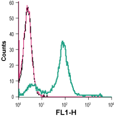
Anti-MCT1 (SLC16A1) (extracellular) Antibody (#AMT-011) is a highly specific antibody directed against an epitope of rat Monocarboxylate transporter 1. The antibody can be used in western blot, immunohistochemistry, and indirect live cell flow cytometry applications. It has been designed to recognize MCT1 from rat, mouse, and human samples.
Applications
Citations
- Western blot of NSC-34 motor neuron-like cell line. Tested in cell lines treated with MCT1/SLC16A1 siRNA.
Gyawali, A. et al. (2022) J. Biomed. Sci. 29, 2.
- Thakur, A. et al. (2020) Sci. Advances. 6, eaaz6119.

