Overview
Application key:
Species reactivity key:

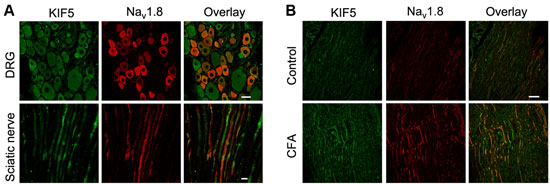
Expression of NaV1.8 in rat DRG and sciatic nerve.Immunohistochemical staining of rat DRG and sciatic nerve using Anti-NaV1.8 (SCN10A) Antibody (#ASC-016). A. NaV1.8 (red) and KIF5B co-localize in DRG and sciatic nerve. B. NaV1.8 (red) and KIF5B expression increases following inflammation induction.Adapted from Su, Y.Y. et al. (2013) with permission of the Society for Neuroscience.
Specifications
- Peptide (C)EDEVAAKEGNSPGPQ, corresponding to amino acid residues 1943-1956 of rat NaV1.8 (Accession Q63554). Intracellular, C-terminus.
Applications
- Rat DRG lysates (1:200). Addition of 0.1% Tween-20 to the blocking and to the antibody solution is recommended.
 Western blot analysis of rat DRG lysates:1. Anti-NaV1.8 (SCN10A) Antibody (#ASC-016), (1:200).
2. Anti-NaV1.8 (SCN10A) Antibody, preincubated with Nav1.8/SCN10A Blocking Peptide (#BLP-SC016).
- Rat dorsal root ganglia (15 µg) (Tan, Z.Y. et al. (2014) J. Neurosci. 34, 7190.).
- Adult rat DRG frozen sections. Human neuromas (1:100) (Black, J.A. et al. (2008) Ann. Neurol. 64, 644.).
- Rat dorsal root ganglia (DRG) (1:500) (Belkouch, M. et al. (2011) J. Neurosci. 31, 18381.).
Scientific Background
Voltage-gated Na+ channels (NaV) are essential for the generation of action potentials and for cell excitability.1 NaV channels are activated in response to depolarization and selectively allow flow of Na+ ions. To date, nine NaV α subunits have been cloned and named NaV1.1-1.9.2-3 The NaV channels are classified into two groups according to their sensitivity to tetrodotoxin (TTX): TTX-sensitive and TTX-resistant channels.4-5 Expression of the α subunit isoform is developmentally and tissue specific.
Two TTX-resistant NaV channels are expressed in dorsal root ganglion (DRG) neurons, NaV1.8 and NaV1.9. The NaV1.8 channel (also called SCN10A, SNS and PN3) is mainly expressed in small-diameter DRG neurons.4-6 TTX-resistant channels have been suggested to play an important role in nociceptive transmission.
Recently, involvement of NaV1.8 in multiple sclerosis (MS) was suggested due to up-regulation of both mRNA and protein in Purkinje cells of MS patients and also in animal models.6
