Overview
- Peptide (C)KTEEEGKTDGEGNAEDGAK, corresponding to amino acid residues 197-215 of rat β2 subunit of voltage-gated Na+ channels (Accession P54900). Intracellular, C-terminus.
 Western blot analysis of rat brain membranes under reducing (lanes 1 and 2) or non-reducing (lanes 3 and 4) conditions:*1,3. Anti-SCN2B (NaVβ2) Antibody (#ASC-007), (1:200).
2,4. Anti-SCN2B (NaVβ2) Antibody, preincubated with SCN2B/Navβ2 Blocking Peptide (#BLP-SC007).
* NaVβ2 is disulfide-linked to the α subunit of voltage-gated Na+ channels (Schmidt, J.W. and Catterall, W.A. (1986) Cell 46, 437.)- Rat brain membranes (1:200-1:1000).
Human cortical lysate (Corbett, B.F. et al. (2013) J. Neurosci. 33, 7020.).
- Rat and mouse brain sections.
Voltage-gated sodium channels (NaV) are essential for the generation of action potentials and for cell excitability.1 To date, nine NaV α subunits have been cloned and named NaV1.1-NaV1.9.2,3 Mammalian sodium channels are heterotrimers, composed of a central, pore-forming α subunit and two auxiliary β-subunits (NaVβ-subunit).4
The NaV β-subunit gene family consists of four members: β1 (SCN1B), β2 (SCN2B), β3 (SCN3B), and β4 (SCN4B) having type I topology, containing an extracellular amino-terminus, a single transmembrane segment, and an intracellular carboxyl-terminus. They modulate channel gating, assembly, and cell surface expression in heterologous cell systems.5
NaV β-subunits are cell adhesion molecules of the Ig superfamily, which interact with extracellular matrix, transmembrane signaling, and cell adhesion molecules.5
In the adult central nervous system and heart, sodium channels are associated with β1- β4 subunits, whereas in adult skeletal muscle they are associated only with the β1 subunit.4,5 This association appears to be a late event in sodium channel biosynthesis.6
Expression of β2-subunits increases in sensory neurons following nerve injury. β2 deficient mice develop less mechanical allodynia in comparison to wild type mice, suggesting a role for β2 subunit in pain sensation.7
Application key:
Species reactivity key:

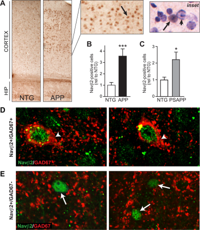
Increased NaVβ2-positive nuclei in cortex of APP mice.A. APP mice exhibit increased numbers of NaVβ2-positive nuclei in cortical regions (arrows). B. Quantification of nuclear NaVβ2 staining in APP and NTG mice, n = 7-9/genotype. C. Quantification of NaVβ2 staining in brain sections from PSAPP mice reveals a significant increase in NaVβ2-positive nuclei also in this mouse model, n 5-7/genotype. D. E. Double-labeling of NaVβ2 and GAD67 in brain sections from APP mice. Confocal images of NaVβ2 and GAD67 immunostaining demonstrate that some NaVβ2-positive nuclei are found in cells that also express GAD67 (D. arrowheads), but others do not express GAD67 (E. arrows). *p>0.05, ***p>0.001.Adapted Corbett, B.F. et al. (2013) with permission of the Society for Neuroscience.
