Overview
- Peptide (C)GKPPSVVSWETRLK, corresponding to amino acid residues 177-190 of human Nectin-1 (Accession Q15223). Extracellular, N-terminus.

 Western blot analysis of rat (lanes 1 and 3) and mouse (lanes 2 and 4) brain lysates:1,2. Anti-Nectin-1/PVRL1 (extracellular) Antibody (#ANR-051), (1:200).
3,4. Anti-Nectin-1/PVRL1 (extracellular) Antibody, preincubated with Nectin-1/PVRL1 (extracellular) Blocking Peptide (#BLP-NR051). Western blot analysis of human Jurkat T-cell leukemia (lanes 1 and 3) and human MEG-01 chronic myelogenous leukemia (lanes 2 and 4) cell lysates:1,2. Anti-Nectin-1/PVRL1 (extracellular) Antibody (#ANR-051), (1:200).
3,4. Anti-Nectin-1/PVRL1 (extracellular) Antibody, preincubated with Nectin-1/PVRL1 (extracellular) Blocking Peptide (#BLP-NR051).
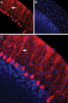
 Expression of Nectin-1 in rat cerebellumImmunohistochemical staining of immersion-fixed, free floating rat brain frozen sections using Anti-Nectin-1/PVRL1 (extracellular) Antibody (#ANR-051), (1:100). A. Nectin-1 staining (red) is apparent in Purkinje neurons and their dendritic tree (arrow). B. Cell nuclei in the same section are visualized with DAPI (blue). C. Merge of the two images.
 Cell surface detection of Nectin-1 by indirect flow cytometry in live intact human MEG-01 megakaryoblastic leukemia cells:___ Cells.
___ Cells + goat-anti-rabbit-FITC.
___ Cells + Anti-Nectin-1/PVRL1 (extracellular) Antibody (#ANR-051), (2.5μg) + goat-anti-rabbit-FITC.- The blocking peptide is not suitable for this application.
 Expression of Nectin-1 in rat PC12 cellsCell surface detection of Nectin-1 in live intact rat PC12 pheochromocytoma cells. A. Extracellular staining of cells with Anti-Nectin-1/PVRL1 (extracellular) Antibody (#ANR-051), (1:100), followed by goat anti-rabbit-AlexaFluor-594 secondary antibody (red). B. Live image of the cells. C. Merge of the two images.
- Rikitake, Y. et al. (2012) J. Cell Sci. 125, 3713.
- Fantin, M. et al. (2013) PLoS One 8, e56897.
- Wakamatsu, K. et al. (2007) J. Biol. Chem. 282, 1817.
- Sözen, M.A. et al. (2001) Nat. Genet. 29, 141.
- Suzuki, K. et al. (2000) Nat. Genet. 25, 427.
Nectins, which were originally identified as virus receptors, are members of the cell-cell adhesion molecule (CAM) family. They are Ca2+-independent immunoglobulin-like CAMs. The nectin family comprises four members, nectin-1, nectin-2, nectin-3 and nectin-4, which are encoded by the PVRL1, PVRL2, PVRL3 and PVRL4 genes, respectively1. In the central nervous system, these cell adhesion molecules aggregate in formations, termed puncta adherentia junctions, which are mechanical adhesive sites that connect pre- and postsynaptic membranes2.
All nectins contain an extracellular region with three immunoglobulin-like loops (one V type and two C2 types), a single membrane-spanning region and a cytoplasmic tail. Nectins directly bind afadin, an F-actin-binding protein, through their cytoplasmic tails1.
Nectin-1 is expressed at cell-cell junctions in human and mouse epidermis. In mice lacking the gene encoding nectin-1 (Pvrl1−/− mice), the expression of loricrin, a differentiation marker and a major component of cornified cell envelopes in the epidermis, is downregulated and newborn pups have a shiny and slightly reddish skin3. Mutations in human PVRL1 are implicated in cleft lip or palate-ectodermal dysplasia syndromes, which includes Zlotogora–Ogur syndrome and Margarita Island ectodermal dysplasia4. Both of these are autosomal recessive disorders that are clinically characterized by unusual facial appearance, dental anomalies, hypotrichosis, palmoplantar hyperkeratosis and onychodysplasia, syndactyly, cleft lip or palate, and in some cases, mental retardation5.
Application key:
Species reactivity key:
Alomone Labs is pleased to offer a highly specific antibody directed against an extracellular epitope of human Poliovirus receptor-related protein 1. Anti-Nectin-1/PVRL1 (extracellular) Antibody (#ANR-051) can be used in western blot, immunocytochemistry and indirect flow cytometry applications. It is specially suited to detect Nectin-1 in live cells. It has been designed to recognize Nectin-1 from rat, mouse and human samples and will recognize all known isoforms of Nectin-1, including the secreted form (also known as isoform gamma).
