Overview
- Peptide (C)DEKHIFSDDSSELTIR, corresponding to amino acid residues 261-276 of rat NCAM-1 (Accession P13596). Extracellular, N-terminus.

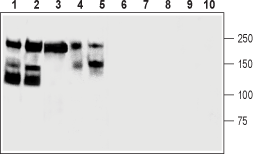
 Western blot analysis of mouse brain (lanes 1 and 6), rat cerebellum (lanes 2 and 7), rat eye (lanes 3 and 8), human SH-SY5Y brain neuroblastoma (lanes 4 and 9) and PC12 rat pheochromocytoma (lanes 5 and 10) cells lysates:1-5. Anti-CD56/NCAM1 (extracellular) Antibody (#ANR-041), (lanes 1-3 1:400 and lanes 4-5 1:200).
6-10. Anti-CD56/NCAM1 (extracellular) Antibody, preincubated with CD56/NCAM1 (extracellular) Blocking Peptide (#BLP-NR041).
 Expression of NCAM1 in rat olfactory bulbImmunohistochemical staining of rat olfactory bulb frozen section using Anti-CD56/NCAM1 (extracellular) Antibody (#ANR-041), (1:300). A. NCAM1 staining (green) appears in glomeruli (G) and is particularly intense in the outer border (arrows). B. Nuclei are stained using DAPI as the counterstain. C. Merged image of A and B panels.
- Johnson, C.P. et al. (2004) Proc. Natl. Acad. Sci. U.S.A. 101, 6963.
- Lanier, L.L. et al. (1991) J. Immunol. 146, 4421.
- Kiss, J.Z. and Muller, D. (2001) Rev. Neurosci. 12, 297.
- Arai, M. et al. (2004) Biol. Psychiatry 55, 804.
- Ćirović, S. et al. (2014) Int. J. Clin. Exp. Pathol. 7, 1402.
Neural cell adhesion molecule 1 (NCAM-1) is a member of a large family of cell-surface glycoproteins and plays a major role during development and controls various functions in the nervous system such as cell migration, neurite growth, axonal guidance, and synaptic plasticity1. NCAM-1 mediates adhesion between cells through homophilic bonds formed between its extracellular domains, which comprise five tandem Ig domains and two juxtamembrane fibronectin type III (Fn III) domains2. There are three major protein isoforms of NCAM-1: 180 kd, 140 kd, and 120 kd. The 180-kd and 140-kd isoforms of NCAM-1 are transmembrane proteins, whereas the 120-kd isoform is linked to the plasma membrane via a glycosyl phosphatidyl-inositol (GPI) lipid anchor3.
NCAM-1 is widely expressed in the central nervous system, skeletal muscle and natural killer cells4.
Several lines of evidence have supported the idea that dysregulation of NCAM-1 isoforms in the brain might be involved in the pathophysiology of neuropsychiatric disorders, particularly bipolar affective disorder5.
Application key:
Species reactivity key:
Anti-CD56/NCAM1 (extracellular) Antibody (#ANR-041) is a highly specific antibody directed against rat Neural cell adhesion molecule 1. The antibody can be used in western blot, live cell imaging, and immunohistochemistry applications. The antibody recognizes an extracellular epitope and is thus ideal for detecting the protein in living cells. It has been designed to recognize NCAM1 from rat, mouse and human samples.
Applications
Citations
- Western blot of brain and developing heart lysates. Tested in NCAM knockout mice.
Delgado, C. et al. (2021) Development. 148, dev199431


