Overview
- Peptide CMIEWPEHPNRTYEK, corresponding to amino acid residues 180-194 of rat NK1 receptor (Accession P14600). 2nd extracellular loop.
- Rat colon sections.
 Expression of NK1 receptor in rat colonImmunohistochemical staining of rat colon paraffin-embedded section using Anti-Neurokinin 1 Receptor (NK1R) (extracellular)-ATTO Fluor-488 Antibody (#ATR-001-AG), (1:100). A. NK1 receptor labeling (green) appears in the tubular glands of the mucosa layer. B. Nuclear staining using DAPI. C. Merge of panels A. and B.
- Human chronic myelogenous leukemia (MEG-O1) living cells 5 µg/5x105 cells.
Substance P (SP), Neurokinin A (NKA) and Neurokinin B (NKB) are all peptides belonging to the Tachykinin protein family. These three peptides, which demonstrate quite a heterogeneity in their distribution, exert their effect via three receptors: Neurokinin 1-3 receptors, members of the G-protein coupled receptor superfamily. However, Neurokinin 1 (NK1) receptor preferentially binds Substance P, Neurokinin 2 (NK2) receptor to NKA, and Neurokinin 3 (NK3) receptor to NKB1.
Neurokinin receptors are distinguished by their seven transmembrane domains, an extracellular N-terminus and a cytosolic C-terminal. An unusual property of these receptors is the presence of introns as part of their structural organization1-3. Tachykinin receptors undergo alternative splicing. For example, NK1 receptor is detected with different C-terminal lengths. The longer receptor isoform is found in the brain whereas the truncated form is mostly detected in the periphery1,4.
Due to the broad expression profile of tachykinin peptides, their respective receptors are also expressed in a similar fashion. NK1 receptor is widely expressed in neurons, endothelial cells, muscle and immune system cells. NK2 receptor is broadly expressed in the periphery and its expression in the brain is quite restricted. NK3 receptor on the other hand is largely expressed in the central nervous system and is also detected in the uterus, skeletal muscle, lung and liver1.
Neurokinin receptors have been found in many pathophysiological indications and have therefore become targets for the development of pharmacological compounds. Such indications include cancer, psychological disorders, migraine and various inflammations, just to name a few5-8.
Application key:
Species reactivity key:
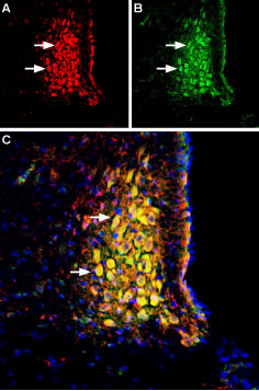
 Multiplex staining of Noradrenaline Transporter (NET) and Neurokinin 1 Receptor (NK1R) in rat brain stem.Immunohistochemical staining of perfusion-fixed frozen rat brain sections using Anti-Noradrenaline Transporter (NET) (extracellular) Antibody (#AMT-002), (1:400) and Anti-Neurokinin 1 Receptor (NK1R) (extracellular)-ATTO Fluor-488 Antibody (#ATR-001-AG), (1:80). A. NET staining (red) in section of rat locus coeruleus. B. NK1 receptor staining (green) in same section. C. Merge of the two images reveals several cells expressing both NET and NK1 receptor (arrows). Cell nuclei are stained with DAPI (blue).
