Overview
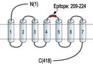
- Peptide EQNRSADGQHAGGLVC corresponding to amino acid residues 209-224 of human NTSR1 (Accession P30989). 2nd extracellular loop.

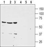
 Western blot analysis of human colon cancer HT-29 (lanes 1 and 4), human lung small cell carcinoma NCI-H526 (lanes 2 and 5) and human breast adenocarcinoma MDA-MB-468 (lanes 3 and 6) cell lysates:1-3. Anti-Neurotensin Receptor 1 (extracellular) Antibody (#ANT-015), (1:200).
4-6. Anti-Neurotensin Receptor 1 (extracellular) Antibody, preincubated with Neurotensin Receptor 1 (extracellular) Blocking Peptide (#BLP-NT015).
 Expression of NTSR1 in human breast cancerImmunohistochemical staining of human breast cancer cells using Anti-Neurotensin Receptor 1 (extracellular) Antibody (#ANT-015), (1:100). Staining (brown color) is specific for epithelium-derivated malignant cells. Hematoxilin is used as the counterstain.
 Expression of NTSR1 in rat brainImmunohistochemical staining of perfusion-fixed frozen rat brain sections using Anti-Neurotensin Receptor 1 (extracellular) Antibody (#ANT-015), (1:100). A. NTSR1 staining (green) appears in cells of the diagonal band region. B. The same section was stained with Choline acetyltransferase (ChAT) (red), a marker of cholinergic neurons. C. Merge of the two images demonstrates expression of NTSR1 in cholinergic neurons. Arrows point at examples of colocalization. Cell nuclei were stained with DAPI (blue).
 Cell surface detection of NTSR1 by indirect flow cytometry in live intact human promyelocytic leukemia HL-60 cells:___ Cells.
___ Cells + goat-anti-rabbit-PE.
___ Cells + Anti-Neurotensin Receptor 1 (extracellular) Antibody (#ANT-015), (2.5μg) + goat-anti-rabbit-PE.- The control antigen is not suitable for this application.
 Expression of NTSR1 in human MCF-7 cellsCell surface detection of NTSR1 in live intact human MCF-7 breast adenocarcinoma cell line. A. Extracellular staining of cells with Anti-Neurotensin Receptor 1 (extracellular) Antibody (#ANT-015), (1:20), followed by Alexa 488-conjugated goat anti-rabbit antibody (green staining). B. Live view of the cells. C. Merge of A and B.
- Vincent, J.P. et al. (1999) Trends Pharmacol. Sci. 20, 302.
- Binder, E.B. et al. (2001) Pharmacol. Rev. 53, 453.
- Souaze, F. et al. (2006) Cancer Res. 66, 6243.
Neurotensin receptor 1 (NTR1) is one of three receptors that mediate the effects of the tridecapeptide neurotensin. Neurotensin is synthesized and secreted from neurons in the central neural system (CNS) and from endocrine cells in the gastrointestinal tract.
NTR1 and NTR2 belong to the 7-transmembrane domain, G-protein coupled receptor (GPCR) superfamily while the third neurotensin receptor NTR3 (also called Sortilin) is a type I membrane protein with a large extracellular domain. Both NTR1 and NTR3 bind neurotensin with high affinity while NTR2 binds it with low affinity.1
NTR1 signals preferentially through Gaq, resulting in the activation phopholipase C and intracellular Ca2+ mobilization, although coupling with Gi/o and to Gs has been observed in some expression systems.
NTR1 is mainly expressed in the brain and gastrointestinal tract. In the brain, NTR1 mediates body temperature regulation, feeding behavior and locomotion regulation. One of the best studied roles of NTR1 is the regulation of dopamine release suggesting that NTR1 can be a suitable target for the development of a new class of anti-psychotics.2
In the periphery, NTR1 expression has been linked to the control of intestine motility and in the stimulation of growth of the intestine mucosa in both physiological and pathological conditions. NTR1 has been shown to be upregulated in several human cancers where it stimulates tumor growth in response to locally produced neurotensin peptide.3
Application key:
Species reactivity key:
Anti-Neurotensin Receptor 1 (extracellular) Antibody (#ANT-015) is a highly specific antibody directed against an epitope of the human NTSR1. The antibody can be used in western blot, immunohistochemistry, live cell imaging, and flow cytometry applications. It has been designed to recognize NTSR1 from human, mouse, and rat samples.
Applications
Citations
- Human B cells (1:200).
Saada, S. et al. (2012) J. Immunol. 189, 5293.
