Overview
- Peptide (C)STDTTDKMQTSLDE, corresponding to amino acid residues 507 - 520 of mouse LRRC4 (Accession Q99PH1). Extracellular, N-terminus.

 Western blot analysis of rat brain synaptosome preparation (lanes 1 and 3) and mouse brain membranes (lanes 2 and 4):1-2. Anti-NGL-2/LRRC4 (extracellular) Antibody (#ANR-162), (1:200).
3-4. Anti-NGL-2/LRRC4 (extracellular) Antibody, preincubated with NGL-2/LRRC4 (extracellular) Blocking Peptide (BLP-NR162). Western blot analysis of human SH-SY5Y neuroblastoma cell line lysates:1. Anti-NGL-2/LRRC4 (extracellular) Antibody (#ANR-162), (1:200).
2. Anti-NGL-2/LRRC4 (extracellular) Antibody, preincubated with NGL-2/LRRC4 (extracellular) Blocking Peptide (BLP-NR162).
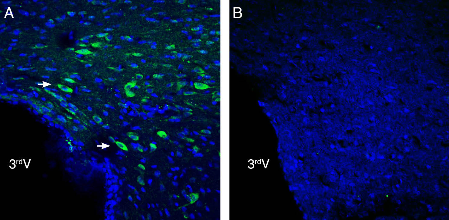
 Expression of NGL-2/LRRC4 in rat hypothalamus.Immunohistochemical staining of perfusion-fixed frozen rat brain sections with Anti-NGL-2/LRRC4 (extracellular) Antibody (#ANR-162), (1:200), followed by goat anti-rabbit-AlexaFluor-488. A. NGL-2/LRRC4 immunoreactivity (green) appears in neurons (arrows). B. Pre-incubation of the antibody with NGL-2/LRRC4 (extracellular) Blocking Peptide (BLP-NR162), suppressed staining. Cell nuclei are stained with DAPI (blue). 3rd V = third cerebral ventricle
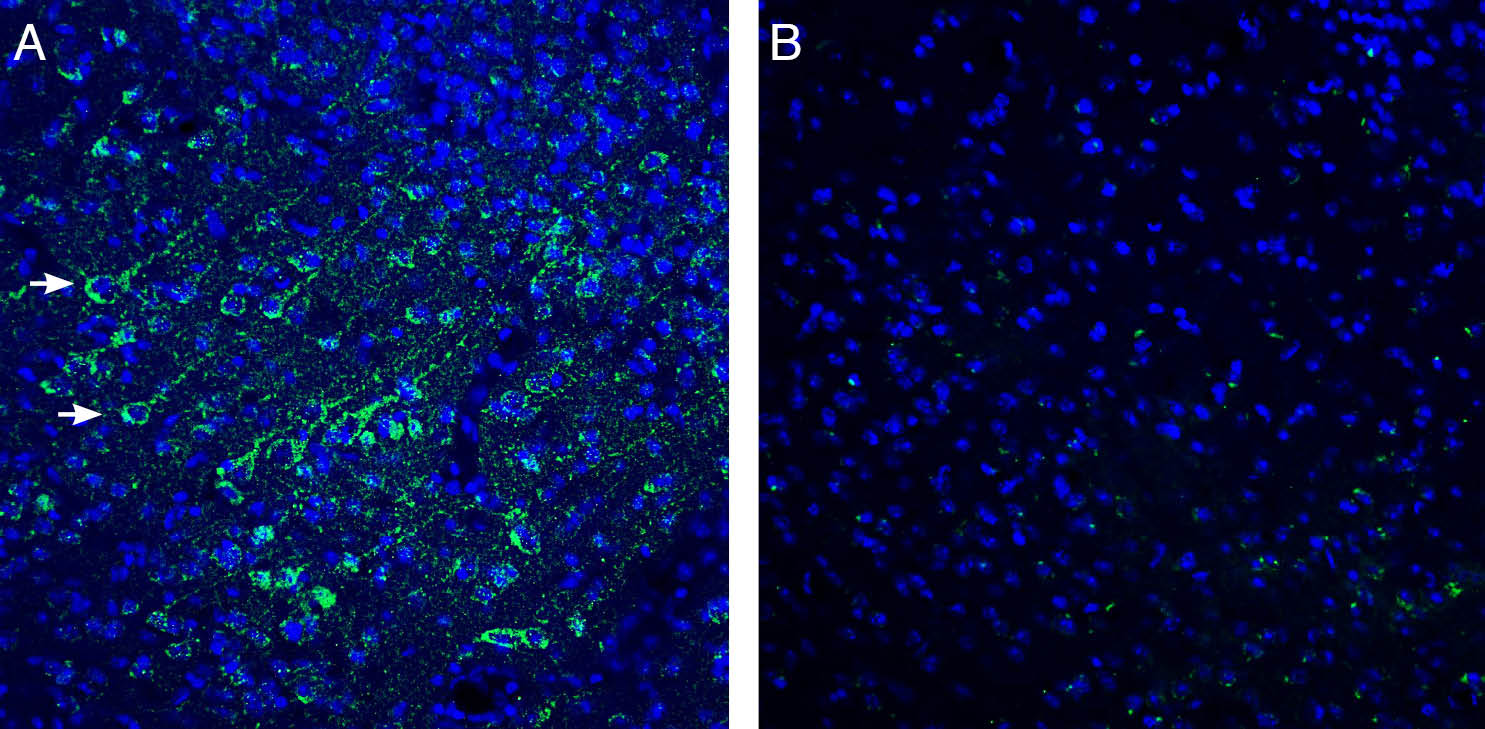
 Expression of NGL-2/LRRC4 in mouse cortex.Immunohistochemical staining of perfusion-fixed frozen mouse brain sections with Anti-NGL-2/LRRC4 (extracellular) Antibody (#ANR-162), (1:200), followed by goat anti-rabbit-AlexaFluor-488. A. NGL-2/LRRC4 immunoreactivity (green) appears in cortical neurons (arrows). B. Pre-incubation of the antibody with NGL-2/LRRC4 (extracellular) Blocking Peptide (BLP-NR162), suppressed staining. Cell nuclei are stained with DAPI (blue).
- Ko, J. (2012) Mol. Cells., 31, 335.
- Zhang, Q. et al. (2005) FEBS. Lett., 4, 3674.
- Li, P. et al. (2014) Mol. Cancer., 19, 1476.
- Um, S.M. et al. (2018) Cell. Rep., 26, 3839.
- Choi, Y. et al. (2019) Front. Mol. Neurosci., 12, 119.
The leucine-rich repeat (LRR) superfamily is a diverse protein family that consist of leucine-rich domains (LRR domains). The LRR domain is known to regulate protein-protein interaction or cell adhesion. Large portion of LRR proteins mediate the differentiation and development of nervous tissues1.
One member of the LRR superfamily is leucine rich repeat containing protein 4 (LRRC4), also known as netrin-G ligand-2 (NGL-2), and serves as a receptor for netrin-G2 (NTNG2). In addition to the LRR domain LRRC4 consist of immunoglobulin (Ig) domain in the extracellular region, a transmembrane domain, and a cytoplasmic region that terminates in a protein-protein interaction domain (PDZ)-binding motif. LRRC4 regulates the formation of excitatory synapses and promotes axon differentiation2.
Functionally, deletion of LRRC4 in mice was found to suppress excitatory synapse development and synaptic transmission. In addition, LRRC4 gene has been identified as tumor suppressor3. In human it has been implicated in autism spectrum disorder (ASD), schizophrenia, and intellectual disability. Moreover, netrin-G2, the presynaptic ligand of LRRC4, is linked to schizophrenia and bipolar disorder4-5.
Application key:
Species reactivity key:
Anti-NGL-2/LRRC4 (extracellular) Antibody (#ANR-162) is a highly specific antibody directed against an extracellular epitope of the mouse protein. The antibody can be used in western blot and immunohistochemistry applications. It has been designed to recognize LRRC4 from rat, mouse and human samples.

