Overview
- Peptide (C)DLEQMERTVDLKD, corresponding to amino acid residues 189-201 of rat nAChRα2 (Accession P12389). Extracellular, N-terminus.

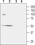
 Western blot analysis of mouse (lanes 1 and 3) and rat (lanes 2 and 4) brain membranes:1,2. Anti-Nicotinic Acetylcholine Receptor α2 (CHRNA2) (extracellular) Antibody (#ANC-002), (1:400).
3,4. Anti-Nicotinic Acetylcholine Receptor α2 (CHRNA2) (extracellular) Antibody, preincubated with Nicotinic Acetylcholine Receptor α2/CHRNA2 (extracellular) Blocking Peptide (#BLP-NC002). Western blot analysis of human SH-SY5Y neuroblastoma cell lysate:1. Anti-Nicotinic Acetylcholine Receptor α2 (CHRNA2) (extracellular) Antibody (#ANC-002), (1:200).
2. Anti-Nicotinic Acetylcholine Receptor α2 (CHRNA2) (extracellular) Antibody, preincubated with Nicotinic Acetylcholine Receptor α2/CHRNA2 (extracellular) Blocking Peptide (#BLP-NC002).
 Expression of nAChRα2 in rat deep cerebellar nucleusImmunohistochemical staining of immersion-fixed, free floating rat brain frozen sections using Anti-Nicotinic Acetylcholine Receptor α2 (CHRNA2) (extracellular) Antibody (#ANC-002), (1:100). Staining reveals expression of nAChRα2 (red) in cells with neuronal outline (arrows point at some examples) in the red nucleus. DAPI is used as the counterstain (blue).
 Expression of nAChRα2 in rat PC12 pheochromocytoma cellsCell surface detection of nAChRα2 in live intact rat PC12 pheochromocytoma cells. A. Extracellular staining of live cells with Anti-Nicotinic Acetylcholine Receptor α2 (CHRNA2) (extracellular) Antibody (#ANC-002), (1:50), followed by goat anti-rabbit-AlexaFluor-594 (red). B. Live image of the cells. C. Merge of the two images.
- Jensen, A.A. et al. (2005) J. Med. Chem. 48, 4705.
- Millar, N.S. and Harkness, P.C. (2008) Mol. Membr. Biol. 25, 279.
- Hogg, R.C. et al. (2003) Rev. Physiol. Biochem. Pharmacol. 147,1.
- Fu, X.W. et al. (2009) Am. J. Respir. Cell. Mol. Biol. 41, 93.
- De Rosa, M.J. et al. (2009) Life Sci. 85, 449.
- Aridon, P. et al. (2006) Am. J. Human Genetics 79, 342.
- Nakauchi, S. et al. (2007) Eur. J. Neurosci. 25, 2666.
Nicotinic acetylcholine receptors (nAChRs) mediate the physiological effects of exogenous nicotine. They also play critical physiological roles throughout the brain and body by mediating cholinergic excitatory neurotransmission, modulating the release of neurotransmitters, and have longer-term effects on gene expression and cellular connections1.
nAChRs are pentameric complexes made up of combinations of a number of different nAChR subunits, which can be classified as α subunits, containing two cysteine residues at positions analogous to Cys192 and Cys193, and non-alpha subunits (‘structural’ subunits), which can be defined as β subunits when they are expressed in the vertebrate nervous system2. There are nine α subunits (α2–α10) and three β subunits (β2, β3, and β4) in the CNS3. Nicotinic receptors are assembled as combinations of α (2-6) and and β (2-4) subunits.
All α subunits are expressed in neuronal cells except for the α1 subunit which is specifically expressed in skeletal muscle. They are also expressed in non-neuronal cells such as bronchial epithelial cells4, as well lymphocytes5.
In humans, a mutant α2 subunit has been identified, which forms nAChRs with increased agonist sensitivity and causes a form of familial epilepsy6. Further, an α2 subunit null mouse model has been used to demonstrate a role for α2 nAChR in nicotine-induced modulation of long-term potentiation in the mouse hippocampal CA1 region, which may underlie some of the cognitive effects of nicotine7.
Application key:
Species reactivity key:
Alomone Labs is pleased to offer a highly specific antibody directed against an extracellular epitope of rat nAChRα2. Anti-Nicotinic Acetylcholine Receptor α2 (extracellular) (CHRNA2) Antibody (#ANC-002) can be used in western blot, immunohistochemistry and immunocytochemistry applications. The antibody is specially suited to recognize nAChRα2 in live cells. It has been designed to recognize nAChRα2 from rat mouse and human samples.
Applications
Citations
- Mouse sample:
Takahashi, T. et al. (2018) Int. J. Mol. Sci. 19, 738.
