Overview
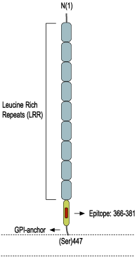
- Peptide (C)GDSPPGNGSGPRHIND corresponding to amino acid residues 366-381 of the human Nogo receptor (Accession Q9BZR6). Extracellular.

 Western blot analysis of rat brain lysate:1. Anti-Nogo Receptor (extracellular) Antibody (#ANT-008), (1:200).
2. Anti-Nogo Receptor (extracellular) Antibody, preincubated with Nogo Receptor (extracellular) Blocking Peptide (#BLP-NT008).
 Expression of Nogo receptor in rat cerebellumImmunohistochemical staining of rat cerebellum using Anti-Nogo Receptor (extracellular) Antibody (#ANT-008). A. NgR (red) appears in Purkinje cells (arrows). B. Staining of Purkinje nerve cells with mouse anti-calbindin D28K (a calcium binding protein, green). C. Confocal merge of NgR and calbindin D28K demonstrates some co-localization of these proteins.
 Expression of Nogo receptor in rat cerebellar granuleCell surface detection of Nogo receptor in live cultured rat cerebellar granule. A. Cells were stained with Anti-Nogo Receptor (extracellular) Antibody (#ANT-008) followed by goat-anti-rabbit AlexaFluor-555 secondary antibody (red). Nuclei were visualized with the cell permeable dye Hoechst 33342 (blue). B. Live view of the same field as in A.
- Fournier, A.E. et al. (2001) Nature 409, 341.
- Prinjha, R. et al. (2000) Nature 403, 383.
- GrandPré, T. et al. (2000) Nature 403, 439.
- Domeniconi, M. et al. (2002) Neuron 35, 283.
- Wang, K.C. et al. (2002) Nature 417, 941.
- Wang, K.C. et al. (2002) Nature 420, 74.
- Mi, S. et al. (2004) Nat. Neurosci. 7, 221.
The Nogo receptor is a leucine-rich repeat (LRR) containing protein with a glycosylphosphatidylinositol (GPI) anchored C-terminus. The receptor was identified on the basis of its ability to bind with high affinity to Nogo-A a member of the reticulon family expressed in the cell membrane of oligodendrocytes.1
Nogo-A attracted attention when it was demonstrated that it is a myelin protein capable of inhibiting axonal growth following nerve injury.2,3 Other myelin proteins also identified as axonal growth inhibitors are the myelin-associated glycoprotein (MAG) a sialic-dependent immunoglobulin-like family member lectin (SIGLEC) and the oligodendrocyte myelin glycoprotein (OmgP) which is a GPI-anchored membrane protein. Remarkably, despite their structural diversity, all three axonal growth inhibitors bind to the Nogo receptor with high affinity.4,5
Since the Nogo receptor is a GPI-anchored protein it was expected that it would require another protein component to transduce the Nogo-A binding information into the responding neurons interior. Indeed, it was shown that the neurotrophin receptor p75 (p75NTR) interacts with Nogo receptor and a third protein termed LINGO-1 to mediate axonal growth inhibition signaling.6,7
Application key:
Species reactivity key:
Alomone Labs is pleased to offer a highly specific antibody directed against an extracellular epitope of the human Nogo receptor. Anti-Nogo Receptor (extracellular) Antibody (#ANT-008) can be used in western blot, immunohistochemistry and live cell imaging applications. It has been designed to recognize NgR from human, mouse and rat samples.
Applications
Citations
- Nakaya, N. et al. (2012) J. Biol. Chem. 287, 37171.
- Rolando, C. et al. (2012) J. Neurosci. 32, 17788.

