Overview
- Peptide CPEPGHKGMDYRDWVRR, corresponding to amino acid residues 16-32 of mouse Orai2 (Accession Q8BH10). Intracellular, N-terminus.

 Western blot analysis of human HL-60 promyelocytic leukemia (lanes 1 and 3) and human Jurkat acute T cell leukemia (lanes 2 and 4) cell lysates:1,2. Anti-Orai2 Antibody (#ACC-061), (1:200).
3,4. Anti-Orai2 Antibody, preincubated with Orai2 Blocking Peptide (#BLP-CC061).
 Expression of Orai2 in rat lungImmunohistochemical staining of paraffin-embedded rat lung sections using Anti-Orai2 Antibody (#ACC-061), (1:100). Strong and specific staining is evident in bronchiolar epithelium (blue) and alveoli walls (red). Hematoxilin is used as the counterstain.
 Expression of Orai2 in mouse cerebellum.Immunohistochemical staining of perfusion-fixed frozen rat brain sections with Anti-Orai2 Antibody (#ACC-061), (1:300), followed by goat anti-rabbit-Alexa-488. A. Orai2 immunoreactivity (green), appears in Purkinje cells (vertical arrows) and in their dendritic tree (horizontal arrows). B. Pre-incubation of the antibody with Orai2 Blocking Peptide (#BLP-CC061), suppressed staining. Cell nuclei are stained with DAPI (blue).
- Eisner, D.A. et al. (2005) Exp. Physiol. 90, 3.
- Chakrabarti, R. and Chakrabarti, R. (2006) J. Cell. Biochem. 99, 1503.
- Feske, S. et al. (2006) Nature 441, 179.
- Pickett, J. (2006) Nature Reviews Mol. Cell Biol. 7, 794.
- DeHaven, W.I. et al. (2007) J. Biol. Chem. 282, 17548.
- Mercer, J.C. et al. (2006) J. Biol. Chem. 281, 24979.
- Gwack, Y. et al. (2007) J. Biol. Chem. 282, 16232.
Cytosolic Ca2+ has long been known to act as a key second messenger in many intracellular pathways including synaptic transmission, muscle contraction, hormonal secretion, and cell growth and proliferation.1,2 The mechanism controlling intracellular Ca2+ level influx, either by the calcium-release-activated Ca2+ channels (CRAC) or from intracellular stores, has become of great interest.
Recently, several key players of the store-operated complex have been identified: the Orai family consisting of three members, Orai1-3, and the STIM family, which consists of two members, STIM1 and STIM2.3 Orai1 (also known as CRACM1) acts as the store-operated calcium channel (SOC) and STIM1 as the endoplasmic reticulum Ca2+ sensor.3,4 Orai1, Orai2, and Orai3 are all capable of forming store-operated channels.5
Co-expresssion of Orai2 and STIM1 was shown to produce currents that appear similar but smaller than those produced by co-expression of Orai1 and STIM1.6
Orai2 transcripts are predominantly expressed in kidney, lung, and spleen, and it appears that Orai2 is the only Orai isoform with multiple transcripts.7
Application key:
Species reactivity key:
Anti-Orai2 Antibody (#ACC-061) is a highly specific antibody directed against an epitope of the mouse protein. The antibody can be used in western blot and immunohistochemistry applications. It has been designed to recognize Orai2 from human, mouse, and rat samples.

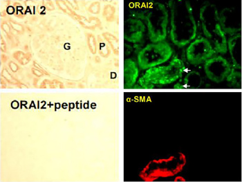
Expression of Orai2 in human kidney.Immunohistochemical staining of human kidney sections using Anti-Orai2 Antibody (#ACC-061). In paraffin-embedded kidney sections, Orai2 is localized to kidney tubules, with stronger staining in the proximal tubules (upper left panel). Antibody preabsorbed with the blocking peptide (supplied with the antibody) was used as control to show the specificity of the antibody (lower left panel). In frozen kidney sections Orai2 staining is observed in renal tubules (upper right panel). Smooth muscle actin staining is shown in red (lower right panel).Adapted from Zeng, B. et al. (2017) Nat. Commun. 8, 1920. with permission of SPRINGER NATURE.
Applications
Citations
- HeLa cell lysate (1:200).
Bittremieux, M. et al. (2017) Cell Calcium 62, 60. - Mouse PA smooth muscle lysate (1:500).
Fernandez, R.A. et al. (2015) Am. J. Physiol. 308, C581.
- Mouse brain sections (1:500).
Kikuta, S. et al. (2019) Front. Cell. Neurosci. 13, 547. - Human kidney sections.
Zeng, B. et al. (2017) Nat. Commun. 8, 1920.
- Mouse brain sections (1:500).
Kikuta, S. et al. (2019) Front. Cell. Neurosci. 13, 547.
- Mitchell, C.M. et al. (2012) J. Neurochem. 122, 1155.
