Overview
- Peptide (C)RNWKRPSEQLEAQH, corresponding to amino acid residues 256-269 of rat OX1R (Accession P56718). 3rd intracellular loop.

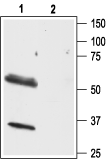
 Western blot analysis of mouse brain lysate:1. Anti-Orexin Receptor 1 Antibody (#AOR-001), (1:200).
2. Anti-Orexin Receptor 1 Antibody, preincubated with Orexin Receptor 1 Blocking Peptide (#BLP-OR001). Western blot analysis of rat brain lysate:1. Anti-Orexin Receptor 1 Antibody (#AOR-001), (1:500).
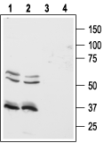
2. Anti-Orexin Receptor 1 Antibody, preincubated with Orexin Receptor 1 Blocking Peptide (#BLP-OR001). Western blot analysis of human Colo-205 (lanes 1 and 3) and HT-29 (lanes 2 and 4) colon cancer cell lines:1,2. Anti-Orexin Receptor 1 Antibody (#AOR-001), (1:500).
3,4. Anti-Orexin Receptor 1 Antibody, preincubated with Orexin Receptor 1 Blocking Peptide (#BLP-OR001).
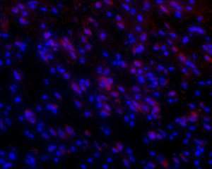
 Expression of OX1R in rat brainLongitudinal frozen section of rat brainstem showing staining (red) in neuronal cell bodies. Slides were incubated overnight at 4°C with Anti-Orexin Receptor 1 Antibody (#AOR-001), (1:50) followed by goat-anti-rabbit-AlexaFluor-555 secondary antibody (1:500). Hoechst 33342 is used as a counterstain (blue).
 Expression of OX1R in rat colonImmunohistochemical staining of paraffin-embedded longitudinal section of rat colon showing mucosa (M), submucosa (SM), and muscularis externa (ME) using Anti-Orexin Receptor 1 Antibody (#AOR-001), (1:100). Note that the stain (red-brown color) is highly specific for absorptive cells in the superior third of the intestinal glands. Immunolabeling was detected using DAB as the chromogen and hematoxilin as the counterstain.
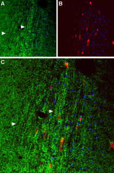
 Expression of OX1R in mouse septumImmunohistochemical staining paraffin-fixed frozen sections using Anti-Orexin Receptor 1 Antibody (#AOR-001), (1:50). A. OX1R (green) appears in axonal processes (right-pointing triangles). B. Parvalbumin (red) appears in septal neurons. Cell nuclei (blue) are visualized with Hoechst 33342. C. Merge of OX1R and parvalbumin suggests that orexinergic innervation covers the entire septal nucleus rather than restricted to individual neurons.
 Expression of OX1R in human colon cancer cell linesImmunocytochemical staining of paraformaldehyde fixed and permeabilized human Colo-205 colon cancer cells A. Cells were stained with Anti-Orexin Receptor 1 Antibody (#AOR-001), (1:200), followed by goat-anti-rabbit-AlexaFluor-555 secondary antibody. B. Live view of the same field as in (A). C. Nuclei were visualized with the cell permeable dye Hoechst 33342 (blue staining).
- Sakurai, T. et al. (1998) Cell. 92, 573.
- Kukkonen, J.P. et al. (2002) Am. J. Physiol. Cell. Physiol. 283, C1567.
- Kirchgessner, A.L. (2002) Endocr. Rev. 23, 1.
- Sakurai, T. (2007) Nat. Rev. Neurosci. 8, 171.
Orexin Receptor 1 (OX1R) (also known as hypocretin receptor 1) is one of two receptors that recognize the peptide neurotransmitters orexin A and orexin B.1 Orexin A and B are 33 and 28 amino acids in length, respectively, and are derived from a common precursor termed prepro-orexin.
OX1R binds orexin A with greater affinity than orexin B (a one order of magnitude difference), while OX2R binds both ligands with similar affinities.2,3
Both OX1R and OX2R belong to the 7-transmembrane domain, G protein-coupled receptor (GPCR) superfamily.
OX1R is thought to transmit signals through the Ga11 class of G proteins, resulting in the activation of phospholipase C with subsequent triggering of the phosphatidylinositol cascade and an influx of extracellular Ca2+, probably through transient receptor potential (TRP) channels.2,3
The physiological functions of the orexin system (OX1R, OX2R, and their ligands) have been a matter of intense research in the last few years.
OX1R is expressed in both the central nervous system and peripheral locations such as gastrointestinal tissues, pancreas, and testis.2 It appears to be involved in the regulation of feeding behavior in rodents since an OX1R antagonist is able to inhibit baseline feeding.2,3
The orexin system has been shown to be involved in the regulation of sleep and wakefulness states and OX1R knockout mice show defects in the regulation of rapid eye movement (REM) sleep, among other phenotypic alterations.4 In addition, the orexin system is involved in regulating autonomic functions such as blood pressure and heart rate, as well as in mechanisms that regulate the reward response in the brain.4
Application key:
Species reactivity key:
Anti-Orexin Receptor 1 Antibody (#AOR-001) is a highly specific antibody directed against an epitope of the rat protein. The antibody can be used in western blot, immunocytochemistry, and immunohistochemistry applications. It has been designed to recognize OX1R from rat, mouse, and human samples.
Applications
Citations
- Rat brain lysate.
Zhou, J.J. et al. (2015) Neuropharmacology 99, 481.
- Rat hypothalamus sections (1:100).
Zhou, J.J. et al. (2015) Neuropharmacology 99, 481.
- Rat isolated rod bipolar cells (RBCs).
Zhang, G. et al. (2018) Neuropharmacology 133, 38.
