Overview
|
Anti-P2X1 Receptor (extracellular) Antibody (#APR-022) is a highly specific antibody directed against an epitope of the human protein. The antibody can be used in western blot, immunocytochemistry, immunohistochemistry, and indirect flow cytometry applications. It has been designed to recognize P2X1 receptor from rat, mouse, and human samples.
Anti-P2X1 Receptor (extracellular)-ATTO Fluor-488 Antibody (#APR-022-AG) is directly labeled with an ATTO-488 fluorescent dye. ATTO dyes are characterized by strong absorption (high extinction coefficient), high fluorescence quantum yield, and high photo-stability. The ATTO-488 label is analogous to the well known dye fluorescein isothiocyanate (FITC) and can be used with filters typically used to detect FITC. Anti-P2X1 Receptor (extracellular)-ATTO Fluor-488 Antibody has been tested in live cell imaging and immunohistochemistry applications and is especially suited for experiments requiring simultaneous labeling of different markers.
Application key:
Species reactivity key:
Applications
 Expression of P2X1 receptor in rat PC12 cellsCell surface detection of P2X1 receptor in intact living PC12 cells. A. Extracellular staining of cells using Anti-P2X1 receptor (extracellular)-ATTO Fluor-488 Antibody (#APR-022-AG), (green), (1:20). B. Merge of extracellular staining with live view of the cells.
 Expression of P2X1 receptor in mouse cerebellumImmunohistochemical staining of mouse cerebellum using Anti-P2X1 Receptor (extracellular)-ATTO Fluor-488 Antibody (#APR-022-AG). A. Most of P2RX1 labeling (green) appears in fine processes in the molecular layer (MOL) and in Purkinje cells. B. Nuclear staining using DAPI. C. Merge of panels A and B.
Specifications
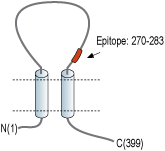
- Peptide CRPIYEFHGLYEEK, corresponding to amino acid residues 270-283 of human P2X1 receptor (Accession P51575). Extracellular loop.

Scientific Background
The P2X receptors belong to the ligand-gated ion channel family and are activated by extracellular ATP.
The structure and function of the P2X receptors, investigated mainly using in vitro models, indicate their involvement in synaptic communication, cell death, and differentiation.
Seven mammalian P2X receptor subtypes (P2X1–P2X7) have been identified and cloned1-3. All P2X receptor subtypes share the same structure of intracellular N- and C-termini two membrane-spanning domains and a large extracellular loop.
All P2X receptor subtypes can assemble to form homomeric or heteromeric functional channels with the exception of P2X6, which only seems to function as part of a heteromeric complex4-9.
The various P2X receptor subtypes show distinct expression patterns. P2X1-6 have been found in the central and peripheral nervous systems, while the P2X7 receptor is predominantly found in cells of the immune system4. The P2X1 receptor is present in smooth muscle, cerebellum, dorsal horn spinal neurons, and platelets where it is suggested to play a regulatory role during in vivo homeostasis and thrombosis3,4,10,11.
- Prasad, M. et al. (2001) J. Physiol. 537, 667.
- Florenzano, F. et al. (2002) Neuroscience 115, 425.
- Ashcroft, F.M. et al. (2000) Ion Channels and Disease Ed 1, p. 405, Academic Press, San Diego.
- Khakh, B.S. et al. (2001) Pharmacol. Rev. 53, 107.
- Ding, Y. et al. (2000) J. Auton. Nerv. Syst. 81, 289.
- Lê, K.T. et al. (1998) J. Neurosci. 18, 7152.
- Robertson, S.J. et al. (2001) Curr. Opin. Neurobiol. 11, 378.
- Dunn, P.M. et al. (2001) Prog. Neurobiol. 65, 107.
- Kim, M. et al. (2001) EMBO J. 20, 6347.
- Burnstock, G. (2001) Trends Pharmacol. Sci. 22, 182.
- Oury, C. et al. (2003) Blood 101, 3969.
