Overview
- Peptide (CS)DPVATSSTLGLQENMRTS, corresponding to amino acid residues 382-399 of rat P2X1 receptor (Accession P47824). Intracellular, C-terminus.

 Western blot analysis of human platelet lysates:1. Anti-P2X1 Receptor Antibody (#APR-001), (1:200).
2. Anti-P2X1 Receptor Antibody, preincubated with P2X1 Receptor Blocking Peptide (#BLP-PR001).
- HEK-293 transfected cells (Agboh, K.C. et al. (2009) Neuropharmacology 56, 230.).
 Expression of P2X1 Receptor in rat hypothalamusImmunohistochemical staining of rat supra-optic nucleus using Anti-P2X1 Receptor Antibody (#APR-001). P2X1 receptor (green) appears in axonal processes (vertical arrows) and cell bodies (horizontal arrows). DAPI is used as the counterstain (blue).
- Mouse mesenteric arteries (Vial, C. and Evans, R.J. (2002) Mol. Pharmacol. 62, 1438.).
- Human Jurkat and CD4+ T cells (Woerhle, T. et al. (2010) Blood 116, 3475.).
- Human skin-resident T cells (MacLeod, A.S. et al. (2014) J. Immunol. 192, 5695.).
- Prasad, M. et al. (2001) J. Physiol. 537, 667.
- Florenzano, F. et al. (2002) Neuroscience 115, 425.
- Ashcroft, F.M. et al. (2000) Ion Channels and Disease Ed 1, p. 405, Academic Press, San Diego.
- Khakh, B.S. et al. (2001) Pharmacol. Rev. 53, 107.
- Ding, Y. et al. (2000) J. Auton. Nerv. Syst. 81, 289.
- Lê, K.T. et al. (1998) J. Neurosci. 18, 7152.
- Robertson, S.J. et al. (2001) Curr. Opin. Neurobiol. 11, 378.
- Dunn, P.M. et al. (2001) Prog. Neurobiol. 65, 107.
- Kim, M. et al. (2001) EMBO J. 20, 6347.
- Burnstock, G. (2001) Trends Pharmacol. Sci. 22, 182.
- Oury, C. et al. (2003) Blood 101, 3969.
The P2X receptors belong to the ligand-gated ion channel family and are activated by extracellular ATP.
The structure and function of the P2X receptors, investigated mainly using in vitro models, indicate their involvement in synaptic communication, cell death, and differentiation.
Seven mammalian P2X receptor subtypes (P2X1–P2X7) have been identified and cloned.1,2,3 All P2X receptor subtypes share the same structure of intracellular N and C-termini two membrane-spanning domains and a large extracellular loop.
All P2X receptor subtypes can assemble to form homomeric or heteromeric functional channels with the exception of P2X6, which only seems to function as part of a heteromeric complex.4-9
The various P2X receptor subtypes show distinct expression patterns. P2X1-6 have been found in the central and peripheral nervous system, while the P2X7 receptor is predominantly found in cells of the immune system.4 The P2X1 receptor is present in smooth muscle, cerebellum, dorsal horn spinal neurons, and platelets where it is suggested to play a regulatory role during in vivo homeostasis and thrombosis.3,4,10,11
Application key:
Species reactivity key:
Alomone Labs is pleased to offer a highly specific antibody directed against an epitope of the rat P2X1 receptor. Anti-P2X1 Receptor Antibody (#APR-001) can be used in western blot, immunoprecipitation, indirect flow cytometry, immunocytochemistry, and immunohistochemistry applications. It has been designed to recognize P2X1 receptor from rat, mouse, and human samples.
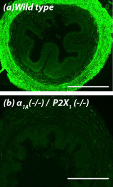
 Knockout validation of Anti-P2X1 Receptor Antibody in mouse vas deferens.Immunohistochemical staining of mouse vas deferens using Anti-P2X1 Receptor Antibody (#APR-001). A. P2X1 receptor staining (green) is detected in the smooth muscle layer of wild-type vas deferens. B. In double α1A-adrenoceptor and P2X1 receptor knockout mice, there is no detection of P2X1 receptor.Adapted from White, C.W. et al. (2013) with permission of the National Academy of Sciences, USA.
Applications
Citations
- Immunohistochemical staining of mouse vas deferens. Tested in KO animals.
White, C.W. et al. (2013) Proc. Natl. Acad. Sci. U.S.A. 110, 20825.
- HEK 293 transfected cell lysate (1:500).
Fryatt, A.G. et al. (2019) J. Gen. Physiol. 151, 146. - Mouse bone lysate and osteoblast MOB-C cell lysate.
Seref-Ferlengez, Z. et al. (2016) PLoS ONE 11, e0155107. - HEK293-TSA 201 transfected cell lysates (1:5000).
Allsopp, R. and Evans, R.J. (2015) J. Biol. Chem. 290, 14556. - Equine palmar digital artery (1:200).
Zamboulis, D.E. et al. (2013) Purinergic Signal. 9, 383. - Rat detrusor cell lysate (1:100).
Uvin, P. et al. (2013) Eur. Urol. 64, 502.
- HEK-293 transfected cells.
Agboh, K.C. et al. (2009) Neuropharmacology 56, 230.
- Rat spinal cord sections.
Vazquez-Villoldo, N. et al. (2014) Glia 62, 171. - Equine palmar digital artery and DRGs (1:200).
Zamboulis, D.E. et al. (2013) Purinergic Signal. 9, 383. - Mouse vas deferens. Also tested in KO animals.
White, C.W. et al. (2013) Proc. Natl. Acad. Sci. U.S.A. 110, 20825. - Mouse mesenteric arteries.
Vial, C. and Evans, R.J. (2002) Mol. Pharmacol. 62, 1438.
- Human Jurkat and CD4+ T cells.
Woerhle, T. et al. (2010) Blood 116, 3475.
- Human eosinophils.
Wright, A. et al. (2016) J. Immunol. 196, 4877. - Human skin-resident T cells.
MacLeod, A.S. et al. (2014) J. Immunol. 192, 5695.
- Kim, M. et al. (2001) J. Biol. Chem. 276, 23262.
- Atkinson, L. et al. (2000) Neuroscience 99, 683.
- Ennion, S. et al. (2000) J. Biol.Chem 275, 29361.
- Gitterman, D.P., and Evans, R.J. (2000) Br. J. Pharmacol. 131, 1561.
- Lewis, C.J. et al. (2000) Br. J. Pharmacol. 129, 124.
- Lewis, C.J., and Evans, R.J. (2000) Br. J. Pharmacol. 131, 1659.
- Vial, C., and Evans, R.J. (2000) Brit. J. Pharmacol. 131, 1489.
