Overview
- Peptide (C)VEKQSTDSGAYSIGH, corresponding to amino acid residues 383-397 of rat P2X3 receptor (Accession P49654). Intracellular, C-terminus.

 Western blot analysis of rat DRG lysates:1. Anti-P2X3 Receptor Antibody (APR-016), (1:200).
2. Anti-P2X3 Receptor Antibody, preincubated with P2X3 Receptor Blocking Peptide (#BLP-PR016).
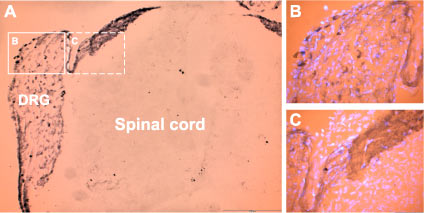
 Expression of P2X3 Receptor in rat DRG neuronsImmunohistochemical staining of rat dorsal root ganglion (DRG) neurons with Anti-P2X3 Receptor Antibody (#APR-016), (A). Cells within the DRG were stained (see solid line frame enlarged (B) as well as fibers and the area of entry of dorsal root into spinal cord (see dashed line frame enlarged (C). DAPI is used as the counterstain (in B and C).
- Fixed mouse trigeminal neurons (1:200) (D'Arco, M. et al. (2007) J. Neurosci. 27, 8190.).
- Human CD4+ and ckit+ cells (Kazakova, R.R. et al. (2011) Bull. Exp. Biol. Med. 151, 33.).
- Florenzano, F. et al. (2002) Neuroscience 115, 425.
- Ashcroft, F.M. et al. (2000) Ion Channels and Disease Ed.1.
- Khakh, B.S. et al. (2001) Pharmacol. Rev. 53, 107.
- Ding, Y. et al. (2000) J. Auton. Nerv. Syst. 81, 289.
- Kage, K. et al. (2002) Exp. Brain Res. 147, 511.
- Prasad, M. et al. (2001) J. Physiol. 573, 667.
- Aoki, Y. et al. (2003) Brain Res. 989, 214.
- Burnstock, G. (2000) Br. J. Anaesth. 84, 476.
The P2X3 receptor belongs to the ligand-gated ion channel P2X receptor family, that consists of seven receptor subtypes named P2X1-P2X7 and is activated by extracellular ATP.1,2,3
All P2X subunits, with the exception of P2X6, can assemble to form homomeric or heteromeric functional channels.4-5 The different P2X receptors show distinct expression patterns. P2X1-6 has been found in the central and peripheral nervous system, while the P2X7 receptor is predominantly found in cells of the immune system. The P2X3 receptor is highly expressed on nociceptive sensory neurons in dorsal root ganglia (DRG) as a homomer or as a heteromer (P2X3/P2X2). ATP released from damaged cells activates the P2X3 receptor to initiate nociceptive signals.6,7 Involvement of ATP in the mechanism of chronic pain has been also suggested.7,8 P2X3 receptor is now becoming a possible target for the development of pain therapeutics.
Application key:
Species reactivity key:
Anti-P2X3 Receptor Antibody (#APR-016) is a highly specific antibody directed against an epitope of the rat protein. The antibody can be used in western blot, indirect flow cytometry, immunohistochemistry, and immunocytochemistry applications. It has been designed to recognize P2X3 receptor from rat, mouse, and human samples.
Applications
Citations
- Rat DRG lysate (1:1000).
Chen, Y. et al. (2016) Mol. Pain 11, 1. - Mouse neuron lysate (1:300).
Gnanasekaran, A. et al. (2013) J. Neurochem. 126, 102.
- Mouse geniculate ganglion (1:500).
Larson, E.D. et al. (2020) eNeuro 7, 0339-19.2020. - Mouse tongue sections (1:300).
Voigt, A. et al. (2015) J. Neurosci. 35, 9717.
- Human ESC NPs (1:100).
Forostyak, O. et al. (2013) Stem Cells Dev. 22, 1506. - Mouse trigeminal neurons (1:200).
Gnanasekaran, A. et al. (2013) J. Neurochem. 126, 102. - Fixed mouse trigeminal neurons (1:200).
D'Arco, M. et al. (2007) J. Neurosci. 27, 8190.
- Mouse geniculate ganglion (1:500).
Larson, E.D. et al. (2020) eNeuro 7, 0339-19.2020.
- Human CD4+ and ckit+ cells.
Kazakova, R.R. et al. (2011) Bull. Exp. Biol. Med. 151, 33.
- Franceschini, A. et al. (2012) BMC Neurosci. doi:10.1186/1471-2202-13-143.
