Overview
- Peptide (C)RDLAGKEQRTLTK, corresponding to amino acid residues 301-313 of rat P2X4 receptor (Accession P51577). Extracellular.

human - 11/13 amino acid residues identical.
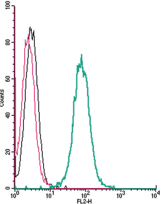
 Cell surface detection of P2X4 Receptor by direct flow cytometry in live intact mouse
BV-2 microglia cells:___ Cells.
___ Cells + Rabbit IgG isotype control-PE.
___ Cells + Anti-P2X4 Receptor (extracellular)-PE Antibody (#APR-024-PE), (2.5µg). Cell surface detection of P2X4 Receptor by direct flow cytometry in live intact human THP-1 monocytic leukemia cells:___ Cells.
___ Cells + Rabbit IgG isotype control-PE.
___ Cells + Anti-P2X4 Receptor (extracellular)-PE Antibody (#APR-024-PE), (2.5µg). Cell surface detection of P2X4 Receptor by direct flow cytometry in live intact human Jurkat T-cell leukemia cells:___ Cells.
___ Cells + Rabbit IgG isotype control-PE.
___ Cells + Anti-P2X4 Receptor (extracellular)-PE Antibody (#APR-024-PE), (2.5µg).
- Prasad, M. et al. (2001) J. Physiol. 537, 667.
- Florenzano, F. et al. (2002) Neuroscience 115, 425.
- Ashcroft, F.M. et al. (2000) Ion Channels and Disease Ed 1, p. 405, Academic Press, San Diego.
- Khakh, B.S. et al. (2001) Pharmacol. Rev. 53, 107.
- Ding, Y. et al. (2000) J. Auton. Nerv. Syst. 81, 289.
- Le, K.T. et al. (1998) J. Neurosci. 18, 7152.
- Robertson, S.J. et al. (2001) Curr. Opin. Neurobiol. 11, 378.
- Dunn, P.M. et al. (2001) Prog. Neurobiol. 65, 107.
- Kim, M. et al. (2001) EMBO J. 20, 6347.
- Inoue, K. et al. (2004) J. Pharmacol. Sci. 94, 112.
The P2X purinergic receptors belong to the ligand-gated ion channel family and are activated by extracellular ATP.
The structure and function of the P2X receptors, which were mainly investigated using in vitro models, indicate their involvement in synaptic communication, cell death, and differentiation.
Seven mammalian P2X receptor subtypes (P2X1–P2X7) have been identified and cloned.1,2,3 All P2X receptor subtypes share the same structure of intracellular N- and C-termini, two membrane-spanning domains and a large extracellular loop.
All P2X subunits can assemble to form homomeric or heteromeric functional channels with the exception of P2X6, which only appears to function as part of a heteromeric complex.4-9
The various P2X receptors show distinct expression patterns. P2X1-6 have been found in the central and peripheral nervous system, while the P2X7 receptor is predominantly found in cells of the immune system.4
The P2X2 receptor subunit has a widespread tissue distribution in autonomic neurons, but it is generally found to be co-expressed with one or more subtypes.
Overexpression of P2X4 was demonstrated in microglia and in the spinal dorsal horn following peripheral nerve injury. It has been suggested that activation of P2X4 along with p38 MAPK is essential for the development of allodynia (pain from a stimulus that doesn't normally elicit pain) following nerve injury. Inhibition of P2X4 expression in spinal microglia has been suggested as a novel therapeutic approach for the treatment of allodynia.10
Application key:
Species reactivity key:
Anti-P2X4 Receptor (extracellular) Antibody (#APR-024) is a highly specific antibody directed against an extracellular epitope of the rat protein. The antibody can be used in western blot, immunocytochemistry and live cell flow cytometry applications. The antibody recognizes an extracellular epitope and can be used to detect the protein in living cells. It has been designed to recognize P2X4 from human, mouse, and rat samples.
Anti-P2X4 Receptor (extracellular)-PE Antibody (#APR-024-PE) is directly conjugated to the Phycoerythrin (PE) fluorophore. This conjugated antibody has been developed to be used in immunofluorescent applications such as direct flow cytometry and live cell imaging.
