Overview
- Peptide (C)KKYKYVEDYEQGLSGEMNQ, corresponding to amino acid residues 370-388 of rat P2X4 receptor (Accession P51577). Intracellular, C-terminus.

 Western blot analysis of rat brain membranes:1. Anti-P2X4 Receptor Antibody (#APR-002), (1:200).
2. Anti-P2X4 Receptor Antibody, preincubated with P2X4 Receptor Blocking Peptide (#BLP-PR002).
- Rat hypothalamus lysate (Jo, Y.H. et al. (2011) J. Biol. Chem. 286, 19993.).
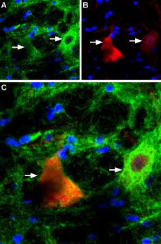
 Expression of P2RX4 in rat brainImmunohistochemical staining of rat brain red nucleus using Anti-P2X4 Receptor Antibody (#APR-002). A. P2RX4 (green) appears in fibers surrounding cell shapes (arrows). B. Calbindin 28K (red) appears in large neurons. C. Merge of P2RX4 and Calbindin 28K suggests variable density of P2RX4 expressing fibers on red nucleus neurons. DAPI is used as the counterstain (blue).
- Mouse brain (1:200) (Jo, Y.H. et al. (2011) J. Biol. Chem. 286, 19993.).
- Human mesangial cells (HMC) (1:200) (Solini, A. et al. (2007) Am. J. Physiol. 292, F1537.).
- NR8383 rat alveolar macrophage cell line (1:100) (Stokes, L. and Surprenant, A. (2009) Eur. J. Immunol. 39, 986.).
- The blocking peptide is not suitable for this application.
- Prasad, M. et al. (2001) J. Physiol. 537, 667.
- Florenzano, F. et al. (2002) Neuroscience 115, 425.
- Ashcroft, F.M. et al. (2000) Ion Channels and Disease Ed 1, p. 405, Academic Press, San Diego.
- Khakh, B.S. et al. (2001) Pharmacol Rev. 53, 107.
- Ding, Y. et al. (2000) J. Auton. Nerv. Syst. 81, 289.
- Le, K.T. et al. (1998) J. Neurosci. 18, 7152.
- Robertson, S.J. et al. (2001) Curr. Opin. Neurobiol. 11, 378.
- Dunn, P.M. et al. (2001) Prog. Neurobiol. 65, 107.
- Kim, M. et al. (2001) EMBO J. 20, 6347.
- Inoue, K. et al. (2004) J. Pharmacol. Sci. 94, 112.
The P2X purinergic receptors belong to the ligand-gated ion channel family and are activated by extracellular ATP.
The structure and function of the P2X receptors, which were mainly investigated using in vitro models, indicate their involvement in synaptic communication, cell death, and differentiation.
Seven mammalian P2X receptor subtypes (P2X1–P2X7) have been identified and cloned.1,2,3 All P2X receptor subtypes share the same structure of intracellular N- and C-termini, two membrane-spanning domains and a large extracellular loop.
All P2X subunits can assemble to form homomeric or heteromeric functional channels with the exception of P2X6, which only appears to function as part of a heteromeric complex.4-9
The various P2X receptors show distinct expression patterns. P2X1-6 have been found in the central and peripheral nervous system, while the P2X7 receptor is predominantly found in cells of the immune system.4
The P2X2 receptor subunit has a widespread tissue distribution in autonomic neurons, but it is generally found to be co-expressed with one or more subtypes.
Overexpression of P2X4 was demonstrated in microglia and in the spinal dorsal horn following peripheral nerve injury. It has been suggested that activation of P2X4 along with p38 MAPK is essential for the development of allodynia (pain from a stimulus that doesn't normally elicit pain) following nerve injury. Inhibition of P2X4 expression in spinal microglia has been suggested as a novel therapeutic approach for the treatment of allodynia.10
Application key:
Species reactivity key:
Anti-P2X4 Receptor Antibody (#APR-002) is a highly specific antibody directed against an epitope of the rat protein. The antibody can be used in western blot, immunocytochemistry, immunohistochemistry, immunoprecipitation, and indirect flow cytometry applications. It has been designed to recognize P2X4 purinergic receptor from rat, mouse, and human samples.

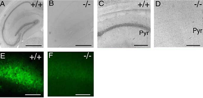
Knockout validation of Anti-P2X4 Receptor antibody in mouse hippocampus.Immunohistochemical staining of mouse hippocampus sections using Anti-P2X4 Receptor Antibody (#APR-002). P2X4 staining is detected in sections from wild type mice (panels A, C, and E) but not in P2X4-/- mice (panels B, D, and F).Adapted from Sim, J.A. et al. (2006) J. Neurosci. 26, 9006. with permission of the Society for Neuroscience.
Applications
Citations
 Expression of P2X4 receptor in lysosomes.Immunocytochemical staining of fixed and permeated COS1 cells using Anti-P2X4 Receptor Antibody (#APR-002). A. P2X4 staining (green) co-localizes with Lamp1 (red), a lysosomal marker. B. Fractionation of COS1 cells shows that P2X4 receptor is detected in the lysosome fraction along with Lamp1. C. Similar results are obtained when P2X4 is heterologously expressed.
Adapted from Huang, P. et al. (2014) with permission of The American Society for Biochemistry and Molecular Biology.
- Mouse DRG lysate. Tested in P2X4-/- mice.
Lalisse, S. et al. (2018) Sci. Rep. 8, 964. - Western blot analysis of mouse brain and liver lysates. Tested in P2X4-/- mice.
Wyatt, L.R. et al. (2014) Neurochem. Res. 39, 1127. - Immunohistochemical staining of mouse hippocampal sections. Tested in P2X4-/- mice.
Sim, J.A. et al. (2006) J. Neurosci. 26, 9006.
- Rat Schwann cells.
Su, W.F. et al. (2019) Glia 67, 78. - Rat DRG lysate (1:300).
Yang, R. et al. (2019) J. Cell. Physiol. 234, 2756. - Mouse DRG lysate. Tested in P2X4-/- mice.
Lalisse, S. et al. (2018) Sci. Rep. 8, 964. - Mouse brain lysate (1:1000).
Areal, L.B. et al. (2017) J. Psychiatr. Res. 91, 57. - Mouse MG6 microglia cell lysate (1:2000).
Hayashi, Y. et al. (2016) Nat. Commun. 7, 11697. - Mouse bone lysate and osteoblast MOB-C cell lysate.
Seref-Ferlengez, Z. et al. (2016) PLoS ONE 11, e0155107. - Rat microglia lysate.
Vazquez-Villoldo, N. et al. (2014) Glia 62, 171. - COS1 cells.
Huang, P. et al. (2014) J. Biol. Chem. 289, 17658. - Mouse brain and liver lysates. Also tested in P2X4-/- mice.
Wyatt, L.R. et al. (2014) Neurochem. Res. 39, 1127. - Human immortalized gingival keratinocytes (HIGK).
Hung, S.C. et al. (2013) PLoS ONE 8, e70210. - Rat Schwann cell lysate (1:500).
Faroni, A. et al. (2013) Cell Death Dis. 4, e743. - Mouse bone marrow-derived dendritic cells (BMDCs) (1:300).
Sakaki, H. et al. (2013) Biochem. Biophys. Res. Commun. 432, 406. - Rat spinal cord lysate.
Lu, W.H. et al. (2013) J. Neurosci. Res. 91, 694. - HEK-293 cells expressing rat P2X4 receptor.
Rokic, M.B. et al. (2013) PLoS ONE 8, e59411.
- Human immortalized gingival keratinocytes (HIGK).
Hung, S.C. et al. (2013) PLoS ONE 8, e70210. - Rat hypothalamus lysate.
Jo, Y.H. et al. (2011) J. Biol. Chem. 286, 19993.
- Mouse vestibular labyrinth sections.
Jeong, J. et al. (2020) Hearing Res. 386, 107860. - Mouse sciatic nerve sections.
Su, W.F. et al. (2019) Glia 67, 78. - Rat DRG sections(1:100).
Yang, R. et al. (2019) J. Cell. Physiol. 234, 2756. - Rat spinal sections.
Vazquez-Villoldo, N. et al. (2014) Glia 62, 171. - Mouse kidney sections (1:2000).
Kim, M.J. et al. (2014) Nephrol. Dial. Transplant. 29, 1350. - Primate retina (1:1000).
Gu, B.J. et al. (2013) FASEB J. 27, 1479. - Rat spinal cord.
Lu, W.H. et al. (2013) J. Neurosci. Res. 91, 694. - Mouse brain (1:200).
Jo, Y.H. et al. (2011) J. Biol. Chem. 286, 19993. - Mouse hippocampal sections. Also tested in P2X4-/- mice.
Sim, J.A. et al. (2006) J. Neurosci. 26, 9006.
- Rat primary microglia cells.
Montilla, A. et al. (2020) Front. Cell. Neurosci. 14, 22. - Rat Schwann cells.
Su, W.F. et al. (2019) Glia 67, 78. - COS1 cells (1:200).
Huang, P. et al. (2014) J. Biol. Chem. 289, 17658. - Mouse striatal mixed glial structures (1:500).
Sorrell, M.E. and Hauser, K.F. (2014) J. Neuroimmune Pharmacol. 9, 233. - Human microglia/monocytic cells.
Vazquez-Villoldo, N. et al. (2014) Glia 62, 171. - Rat Schwann cells (1:1000).
Faroni, A. et al. (2013) Cell Death Dis. 4, e743. - Rat primary microglia.
Lu, W.H. et al. (2013) J. Neurosci. Res. 91, 694. - Human mesangial cells (HMC) (1:200).
Solini, A. et al. (2007) Am. J. Physiol. 292, F1537.
- Mouse vestibular labyrinth sections.
Jeong, J. et al. (2020) Hearing Res. 386, 107860. - Rat primary microglia cells.
Montilla, A. et al. (2020) Front. Cell. Neurosci. 14, 22.
- NR8383 rat alveolar macrophage cell line (1:100).
Stokes, L. and Surprenant, A. (2009) Eur. J. Immunol. 39, 986.
- Thompson, K.E. et al. (2013) FASEB J. 27, 1772.
- Roberts, V.H. et al. (2007) Placenta 28, 270.
- Cavaliere, F. et al. (2004) J. Cereb. Blood Flow Metab. 24, 392.
- Kim, M. et al. (2001) J. Biol. Chem. 276, 23262.
- Rubio, M.E. and Soto, F. (2001) J. Neurosci. 21, 641.
- Gitterman, D.P. and Evans, R.J. (2000) Br. J. Pharmacol. 131, 1561.
- Lewis, C.J. and Evans, R.J. (2000) Br. J. Pharmacol. 131, 1659.
- Vial, C. and Evans, R.J. (2000) Br. J. Pharmacol. 131, 1489.
