Overview
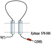
- Peptide (C)KIRKEFPKTQGQYSGFKYPY, corresponding to amino acid residues 576-595 of rat P2X7 receptor (Accession Q64663). Intracellular, C-terminus.

 Multiplex staining of IBA1/AIF1 and P2X7 in rat brain.Immunohistochemical staining of rat corpus callosum (CC) free floating frozen sections using Anti-IBA1/AIF1 Antibody (#ACS-010), (1:1000) and Anti-P2X7 Receptor-ATTO Fluor-550 Antibody (#APR-004-AO) (1:60). A. IBA1/AIF1 immunoreactivity (green) appears in microglia (arrows). B. P2X7 immunostaining (red) appears in microglia (up-pointing arrows) and in other cell types in the corpus callosum (down-pointing arrows). C. Merged image of panels A and B demonstrates partial colocalization of both proteins. Nuclei are demonstrated using DAPI as the counterstain (blue).
 Expression of P2RX7 in rat hippocampusImmunohistochemical staining of rat hippocampus frozen section using Anti-P2X7 Receptor-ATTO Fluor-550 Antibody (#APR-004-AO), (1:60). A. P2X7 staining (red) appears in the CA1 pyramidal (Pyr) and stratum radiatum (SR) layers in cells with glial morphology (arrows). B. DAPI is used as the counterstain (blue). C. Merged image of A and B.
 Multiplex staining of VGLUT2 and P2X7 Receptor in rat spinal cordImmunohistochemical staining of perfusion-fixed frozen rat spinal cord sections using Anti-VGLUT2 Antibody (#AGC-036), (1:600) and Anti-P2X7 Receptor-ATTO Fluor-550 Antibody (#APR-004-AO), (1:100). A. Vesicular Glutamate Transporter 2 labeling followed by goat-anti-rabbit-Alexa-488 (green). B. The same section labeled for P2X7 Receptor (orange). C. Merge of A and B demonstrates partial co-localization of VGLUT2 and P2X7 Receptor in dorsal horn and in lateral column (L. Col., arrow). Cell nuclei were stained with DAPI (blue).
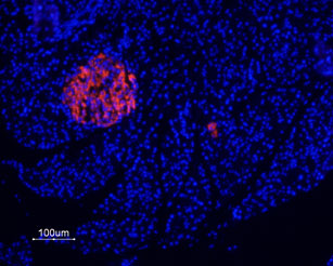
 Expression of P2X7 Receptor in rat pancreasImmunohistochemical staining of rat paraffin embedded endocrine and exocrine pancreas sections using Anti-P2X7 Receptor-ATTO Fluor-550 Antibody (#APR-004-AO), (1:20), (red). Staining is highly specific for endocrine cells of the Isle of Langerhans. Hoechst 33342 is used as the counterstain (blue).
- Mouse spinal cord sections (1:20) (Apolloni, S. et al. (2013) Hum. Mol. Genet. 22, 4102.).
- Ding, Y. et al. (2000) J. Auton. Nerv. Syst. 81, 289.
- Kim, M. et al. (2001) EMBO J. 20, 6347.
- Chizh, B.A. and Illes, P. (2001) Pharmacol. Rev. 53, 553.
The P2X7 purinergic receptor is a member of the ionotropic P2X receptor family that is activated by ATP. To date, this family consists of seven receptor subtypes, named P2X1-P2X7, all of which have been cloned.
The various P2X receptors show distinct expression patterns. P2X1-6 receptors have been found in the central and peripheral nervous system, while the P2X7 receptor is found in cells of the immune system, particularly in antigen-presenting cells, and microglia.
The P2X7 receptor mediates the release of proinflammatory cytokines and stimulation of transcription factors and may also play an important role in apoptosis.1-3
Application key:
Species reactivity key:
Anti-P2X7 Receptor Antibody (#APR-004) is a highly specific antibody directed against an epitope of the rat protein. The antibody can be used in western blot, immunoprecipitation, immunocytochemistry, and immunohistochemistry applications. It has been designed to recognize P2X7 purinergic receptor from rat, mouse, and human samples.
Anti-P2X7 Receptor-ATTO Fluor-550 Antibody (#APR-004-AO) is directly labeled with an ATTO-550 fluorescent dye. ATTO dyes are characterized by strong absorption (high extinction coefficient), high fluorescence quantum yield, and high photo-stability. The ATTO-550 fluorescent label is related to the well known dye Rhodamine 6G and can be used with filters typically used to detect Rhodamine. Anti-P2X7 Receptor-ATTO Fluor-550 Antibody has been tested in immunohistochemistry applications and is specially suited to experiments requiring simultaneous labeling of different markers.

Expression of P2X7 purinergic receptor in mouse spinal cordImmunohistochemical staining of mouse spinal cord sections (L3–L5) from SOD1-G93A mice were double-immunostained either with anti-Iba-1, NeuN or GFAP (green) and Anti-P2X7 Receptor-ATTO Fluor-550 Antibody (#APR-004-AO), (red). P2X7 is present only in microglia cells as shown in the merged panel (yellow). Scale bar = 20 μm, insets = 50 μm.Adapted from Apolloni, S. et al. (2013) Hum. Mol. Genet. 22, 4102. with permission of Oxford University Press.
Applications
Citations
- Western blot analysis of mouse neutrophil lysate using #APR-004. Tested in P2X7-/- mice.
Karmakar, M. et al. (2016) Nat. Commun. 7, 10555. - Western blot analysis of mouse GL261 glioma cell lysate using #APR-004. Tested on siRNA treated cells.
Gehring, M.P. et al. (2015) Int. J. Biochem. Cell Biol. 68, 92. - Immunocytochemical staining of mouse microglial cells with #APR-004 (1:600). Tested on P2X7-/- cells.
Fischer, W. et al. (2014) Purinergic Signal. 10, 313. - Western blot analysis of mouse lumbar spinal cord protein lysates using #APR-004 (1:500). Tested in P2X7-/- mice.
Apolloni, S. et al. (2013) Hum. Mol. Genet. 22, 4102.
- Rat brain sections.
Kuan, Y.H. et al. (2015) Neurobiol. Dis. 78, 134. - Mouse spinal cord sections (1:20).
Apolloni, S. et al. (2013) Hum. Mol. Genet. 22, 4102.
