Overview
- Peptide SDEYLRSYFIYSMC, corresponding to amino acid residues 207-220 of human P2RY1 (Accession P47900). 2nd extracellular loop.

 Western blot analysis of rat brain (lanes 1 and 4), Jurkat (lanes 2 and 5) and K-562 (lanes 3 and 6) lysates:1-3. Anti-P2Y1 Receptor (extracellular) Antibody (#APR-021), (1:200).
4-6. Anti-P2Y1 Receptor (extracellular) Antibody, preincubated with P2Y1 Receptor (extracellular) Blocking Peptide (#BLP-PR021).
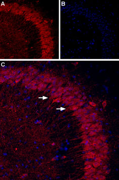
 Expression of P2RY1 in mouse hippocampusImmunohistochemical staining of mouse hippocampal CA3 region using Anti-P2Y1 Receptor (extracellular) Antibody (#APR-021). A. P2RY1 staining (red) appears in the pyramidal layer (arrows). B. Nuclei staining using DAPI as the counterstain (blue). C. Merged images of panels A and B.
 Expression of P2RY1 in rat PC12 cellsCell surface detection of P2RY1 in intact living rat pheochromocytoma PC12 cells. A. Extracellular staining of cells using Anti-P2Y1 Receptor (extracellular) Antibody (#APR-021), (1:50) followed by goat anti-rabbit-AlexaFluor-594 secondary antibody (red). B. Merge of extracellular staining with live view of the cells.
- Wang, L. et al. (2002) J. Cardiovas. Pharmacol. 40, 841.
- Queiroz, G. et al. (2003) J. Pharmacol. Exp. Ther. 307, 809.
- Ralevic, V. and Burnstock, G. (1998) Pharmacol. Rev. 50, 413.
- Abbracchio, M.P. et al. (2003) Trends Pharmacol. Sci. 24, 52.
- Jin, J. et al. (1998) J. Biol. Chem. 273, 2030.
The P2Y receptors belong to the G-protein coupled receptors superfamily. P2Y receptors mediate the actions of the extracellular nucleotides (ATP, ADP, UTP and UDP) and regulate several physiologic responses, among them, cardiac function, platelet aggregation and SMC proliferation.1
The mammalian P2Y family currently includes eight functional receptors; P2Y1, P2Y2, P2Y4, P2Y6, P2Y11, P2Y12, P2Y13, and the UDP-glucose receptor, now renamed P2Y14.2-4
P2Y1 receptor is abundantly expressed in a number of tissues. In platelets, P2Y1 receptor is co-expressed with P2Y12 receptor, responsible for the initial shape change, aggregation, and rise in intracellular Ca2+ upon activation in response to ADP.5
Application key:
Species reactivity key:
Anti-P2Y1 Receptor (extracellular) Antibody (#APR-021) is a highly specific antibody directed against an extracellular epitope of the human P2RY1. The antibody can be used in western blot, immunohistochemistry, indirect flow cytometry and live cell imaging applications. It has been designed to recognize P2RY1 from mouse, rat and human samples.
Applications
Citations
- Mouse DRG Sections (1:650).
Mitchell, R. et al. (2019) Mol. Neurobiol. 56, 5917.
- Mouse DRG Sections (1:650).
Mitchell, R. et al. (2019) Mol. Neurobiol. 56, 5917.
- Mouse primary microglia culture.
Fukumoto, Y. et al. (2018) J. Cereb. Blood Flow Metab. 39, 2144.

