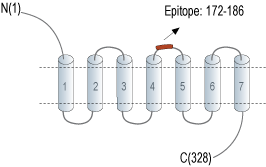
Overview
- Peptide (C)RNRTV(S)YDLSPPILS, corresponding to amino acid residues 172 - 186 of mouse P2RY6 (Accession Q9ERK9). Extracellular, 2nd loop.

 Cell surface detection of P2Y6 by direct flow cytometry in live intact mouse J774 macrophage cells:___ Cell alone
___ Cells + Rabbit IgG isotype control-PE
___ Cells + Anti-P2Y6 Receptor (extracellular)-PE Antibody (APR-106-PE), 2.5µg. Cell surface detection of P2Y6 by direct flow cytometry in live intact mouse BV-2 microglia cells:___ Cell alone
___ Cells + Rabbit IgG isotype control-PE
___ Cells + Anti-P2Y6 Receptor (extracellular)-PE Antibody (APR-106-PE), 2.5µg.
- Communi, D. et al. (2000) Cell Signal. 12, 351.
- Filippov, A.K. et al. (1999) Br. J. Pharmacol. 126, 1009
- Chang, K. et al. (1995) J. Biol. Chem. 270, 26152.
- Malmsjo, M. et al. (2003) B.M.C. Pharmacol. 3, 4.
The nucleotide receptor P2Y6 binds UDP, and is a member of the G-protein coupled receptor superfamily1,3.
P2Y6 agonists induce an inositol phosphate/Ca2+ response that is insensitive to pertussis toxin inhibition. P2Y6 is coupled to phospholipase C via Gq, and is also coupled to inhibition of N-type Ca2+ and M-type K+ channels1,2.
P2Y6 mRNA is expressed in various rat tissues, including lung, stomach, intestine, spleen, heart, and aorta. P2Y6 was reported to mediate contractions in human cerebral arteries4.
Application key:
Species reactivity key:
Anti-P2Y6 Receptor (extracellular) Antibody (#APR-106) is a highly specific antibody directed against an extracellular epitope of the mouse protein. The antibody can be used in western blot, immunohistochemistry, and indirect flow cytometry applications. It has been designed to recognize P2RY6 from human, rat, and mouse samples.
Anti-P2Y6 Receptor (extracellular)-PE Antibody (#APR-106-PE) is directly conjugated to the Phycoerythrin (PE) fluorophore. This conjugated antibody has been developed to be used in immunofluorescent applications such as direct flow cytometry and live cell imaging.
