Overview
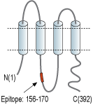
- Peptide (C)RLVQHMLKIRQKSSD, corresponding to amino acid residues 156-170 of human PANX3 (Accession Q96QZ0). Intracellular loop.

 Western blot analysis of rat (lanes 1 and 3) and mouse (lanes 2 and 4) testis lysates:1,2. Anti-Pannexin 3 Antibody (#ACC-233), (1:200).
3,4. Anti-Pannexin 3 Antibody, preincubated with Pannexin 3 Blocking Peptide (#BLP-CC233). Western blot analysis of rat skeletal muscle lysate:1. Anti-Pannexin 3 Antibody (#ACC-233), (1:200).
2. Anti-Pannexin 3 Antibody, preincubated with Pannexin 3 Blocking Peptide (#BLP-CC233).
- Panchin, Y. et al. (2000) Curr. Biol. 10, R473.
- Sosinsky Gina, E. et al. (2011) Channels 5, 193.
- Penuela, S. et al. (2007) J. Cell Sci. 120, 3772.
Pannexins are a class of membrane channels similar to the invertebrate Connexin family of gap channel proteins. There are currently three known types of pannexin channels and the exact role of these channels is yet to be fully discerned1.
A recent study suggests that despite similarity to the connexin family these channels are not gap-junctions or “hemichannels” as described in some publications but fully operational channels. These conclusions were derived because pannexins, unlike gap-junctions, are found to be expressed and functional in single membranes facing the extracellular space and not as intercellular membranes in appositional cells.
Pannexins are comprised of four transmembrane segments, two extracellular loops and both the amino and carboxy termini are facing the cytosol. Pannexins also have four conserved extracellular loop cysteines resulting in the possibility of two intra-pannexin disulfide bonds. Pannexin 3 has a glycosylation site at N254 and N71. Pannexin membrane channels are formed by 6-8 monomers and while pannexins 3's exact oligomeric number has not been determined yet it is expected to be 6 with a greater similarity to Pannexin 1 than 22.
There are two known subtypes of Panx3 in mice with different molecular weight and distribution. The first, weighing 43 kDa is mostly expressed in cartilage skin tissues and also in low levels in heart ventricle tissue. The second, weighing 70 kDa, and possibly functioning as a dimer is detected in lung, liver, thymus and spleen tissues3.
Since full understanding of pannexin 3 structure, expression and role in physiological and pathological tissues is yet to be obtained there is a definite need for further research.
Application key:
Species reactivity key:
Alomone Labs is pleased to offer a highly specific antibody directed against an epitope of human Pannexin 3. Anti-Pannexin 3 Antibody (#ACC-233) can be used in western blot analysis. It was designed to recognize PANX3 from rat, mouse and human samples.
