Overview
- Peptide (C)KVRVDGSQDAQLYE, corresponding to amino acid residues 440-453 of mouse PLXNA3 (Accession P70208). Extracellular, N-terminus.

 Western blot analysis of rat (lanes 1 and 3) and mouse (lanes 2 and 4) brain membranes:1,2. Anti-Plexin-A3 (extracellular) Antibody (#APR-093), (1:200).
3,4. Anti-Plexin-A3 (extracellular) Antibody, preincubated with Plexin-A3 (extracellular) Blocking Peptide (#BLP-PR093). Western blot analysis of human THP-1 monocytic leukemia cell line lysate:1. Anti-Plexin-A3 (extracellular) Antibody (#APR-093), (1:200).
2. Anti-Plexin-A3 (extracellular) Antibody, preincubated with Plexin-A3 (extracellular) Blocking Peptide (#BLP-PR093).
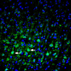
 Expression of Plexin-A3 in mouse parietal cortexImmunohistochemical staining of perfusion-fixed frozen mouse brain sections with Anti-Plexin-A3 (extracellular) Antibody (#APR-093), (1:200), followed by goat-anti-rabbit-AlexaFluor-488. PLXNA3 staining (green) appears in pyramidal neurons (arrows). Cell nuclei are stained with DAPI (blue).
- Zhang, L. et al. (2015) PLoS One 10, e0121513.
- Barton, R. et al. (2015) PLoS One 10, e0116368.
Plexins are type 1 transmembrane (TM) receptors acting as key regulatory proteins in a wide variety of developmental, regenerative and pathological processes. They are involved in guidance and motility of vascular, lymphatic vessel, and neuron growth, and in cancer metastasis.
Plexin family includes 9 proteins: Plexin-A1-4, -B1-3, -C1 and -D1. Class A Plexins interact with the secreted class 3 Semaphorins, and require Neuropilins (nrps) as co-receptors.
Plexin protein signaling results in cytosolic GTPase-activating
protein activity including direct interactions with Rho GTPases and transient/catalytic interactions with Ras GTPases1,2.
Plexin structure consists of an extracellular semaphorin domain, three Plexin-Semaphorin-Integrin (PSI) domains, and three immunoglobulin, Plexin, and transcription factor (IPT) domains. In addition, a single-spanning transmembrane domain, and a cytosolic region (CYTO) homologous with Ras GTPase-activating proteins (GAPs)2. The transmembrane region of Plexin-A3 is characterized by poly-glycine usually used for close helix-helix packing and aromatic residues that distort helix conformation and association. Plexin-A family members only have 22 residues spanning the membrane1.
Mutations in Plexins have been reported in melanoma, lung, breast, pancreatic, and prostate cancers, strongly suggesting that their signaling may play a role in cancer development. Plexins are widely expressed in the nervous system and in the developing cerebral cortex2.
Application key:
Species reactivity key:
Anti-Plexin-A3 (extracellular) Antibody (#APR-093) is a highly specific antibody directed against an epitope of the mouse protein. The antibody can be used in western blot, immunohistochemistry and indirect live cell flow cytometry. The antibody recognizes an extracellular epitope, and can potentially be used for detecting the protein in living cells. It has been designed to recognize PLXNA3 from rat, mouse, and human samples.

