Overview
- Peptide CDRILNKEGGIVPFKTK, corresponding to amino acid residues 601-617 of rat PMCA4 (Accession Q64542). 2nd intracellular loop.
- Rat stomach and lung lysates (1:200-1:1000).
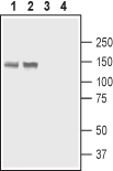
 Western blot analysis of rat stomach lysate (lanes 1 and 3) and rat lung membranes (lanes 2 and 4):1,2. Anti-PMCA4 Antibody (#ACP-004), (1:200).
3,4. Anti-PMCA4 Antibody, preincubated with PMCA4 Blocking Peptide (#BLP-CP004).
- Rat brain sections (1:300).
Plasma membrane calcium pumps (PMCA) are part of the P2 subfamily of P-type primary ion transport ATPases. PMCAs are found in all eukaryotic cells and serve as a major high-affinity transporter for Ca2+ in the plasma membrane.
PMCA4, as other PMCA pumps, is predicted to have 10 membrane spanning domains with the -NH2 and -COOH termini both located on the cytosolic side of the membrane. Most of the protein is facing the cytosol and is comprised of three main parts - the intracellular loop between transmembrane segments 2 and 3, the large unit between membrane-spanning domains 4 and 5, and an extended “tail” following the last transmembrane domain.
PMCA4 is regulated by various regulatory mechanisms including Ca2+-Calmodulin and acidic phospholipids. Calmodulin binds to a region in the -COOH terminal located about 40 residues downstream from the last transmembrane domain. In the absence of calmodulin, this region acts as an auto-inhibitory domain. Acidic phospholipids are strong activators of PMCA4. They reduce the Michaelis-Menten constant for Ca2+ of the enzyme to roughly 0.3 µM compared with 0.4–0.7 µM in the case of calmodulin activation. Activation by phospholipids makes the PMCA insensitive to calmodulin thus suggesting that their effect is not additive1.
PMCA4 deficiency does not affect embryonic development in mice. PMCA4 knockout mice are indistinguishable from wild type mice in terms of their behavior, appearance and weight. PMCA4 is highly expressed in the sperm tail and deletion of PMCA4 causes severely impaired sperm motility in mice2.
