Overview
- Peptide (C)DEDQKVRPNEENNKDAD, corresponding to amino acid residues 72-88 of human BDNF (precursor) (Accession P23560). Pro-domain of the BDNF protein.

 Western blot analysis of rat glioma C6 (1, 3) or human neuroblastoma SH-SY5Y (2, 4) cell lysate:1,2. Anti-proBDNF Antibody (#ANT-006), (1:200).
3,4. Anti-proBDNF Antibody, preincubated with proBDNF Blocking Peptide (#BLP-NT006). Western blot analysis using Anti-proBDNF Antibody (#ANT-006), (1:400):1. Recombinant human proBDNF protein (#B-257), (20 ng).
2. Recombinant human proNGF protein (#N-280), (200 ng).
3. Recombinant proNT-3 (200 ng).
4. Recombinant human BDNF protein (#B-250), (200 ng).
5. Native mouse NGF 2.5S protein (>95%) (#N-100), (200 ng).
6. Recombinant human Neurotrophin-3 (NT-3) protein (#N-260), (200 ng).
- Rat hippocampal neurons (Danelon, V. et al. (2016) Mol. Cell. Neurosci. 75, 81.).
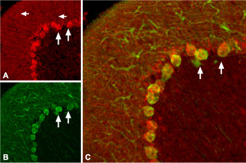
 Expression of proBDNF in rat cerebellumImmunohistochemical staining of rat cerebellum with Anti-proBDNF Antibody (#ANT-006). proBDNF (red) appears in Purkinje cell bodies (vertical arrows) and in astrocytic processes (horizontal arrows in A) but not in Purkinje neuronal dendrites stained for calbindin D28k (green, in B) in the same brain section. C. Confocal merge of proBDNF and CBD28K demonstrates the restriction of proBDNF to Purkinje cell body.
 Multiplex staining of BDNF and proBDNF in rat hippocampusImmunohistochemical staining of rat hippocampal dentate gyrus perfusion-fixed frozen sections using Guinea pig Anti-BDNF Antibody (#ANT-010-GP), (1:300) and Anti-proBDNF Antibody (#ANT-006), (1:200). A. BDNF staining (red) appears in interneuron outlines (arrows) in the hilus (H) region and in the granule layer (G). B. proBDNF staining (green) in the same section appears in interneuron outlines (arrows) in the hilus (H) region and in the granule layer (G). C. Merge of panel A and panel B shows extensive co-localization of the two proteins.
- Lee R. et al. (2001) Science 294,1945.
- Egan, M.F. et al. (2003) Cell 112, 257.
- Pang, P.T. et al. (2004) Science 306, 487.
- Michalski, B. et al. (2003) Mol. Brain Res. 111, 148.
Brain derived neurotrophic factor (BDNF) is a member of the neurotrophin family of growth factors that includes nerve growth factor (NGF), neurotrophin-3 (NT-3) and neurotrophin-4/5 (NT-4/5).
All neurotrophins are synthesized as preproneurotrophin precursors that are subsequently processed within the intracellular transport pathway to yield proneurotrophins that are further processed to generate the mature form. The mature form of BDNF is a non-covalent stable homodimer that can be secreted in both constitutive and regulated pathways.
Until recently, the functional role of the neurotrophin prodomains were thought to include assistance in the correct folding of the mature protein and the sorting of the neurotrophins into the constitutive or regulated secretory pathway. However, a growing body of evidence suggests that the uncleaved proneurotrophin precursors can be secreted from cells and that they may mediate different biological functions.
The functional importance of the prodomain of BDNF was recently demonstrated in a study showing that a polymorphism that replaces valine for methionine at position 66 of the prodomain, is associated with memory defects and abnormal hippocampal function in humans. Another recent study showed that the regulated extracellular cleavage of proBDNF to mature BDNF by plasmin is necessary for establishing late-phase long-term potentiation (L-LTP), a process that involves long-lasting changes in the structure and function of hippocampal synapses. Finally, proBDNF was shown to be decreased in the brains of patients suffering from Alzheimer’s disease.
Mature BDNF binds to the specific tyrosine kinase receptor TrkB and to p75NTR, a member of the TNF receptor superfamily. proBDNF can similarly bind both receptors although it appears to have a greater affinity for the p75NTR receptor.
Application key:
Species reactivity key:
Anti-proBDNF Antibody (#ANT-006) is a highly specific antibody directed against the prodomain region of human proBDNF. The antibody can be used in western blot, immunoprecipitation, and immunohistochemistry applications. It has been designed to recognize proBDNF from human, mouse, and rat samples. The antibody does not cross react with mature BDNF, pro- and mature NGF or mature NT-3.

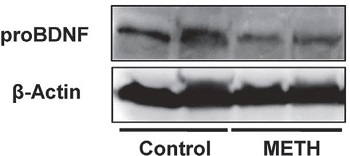
Expression of proBDNF in mouse prefrontal brain cortex.Western blot analysis of mouse prefrontal brain cortex lysates using Anti-proBDNF Antibody (#ANT-006). proBDNF expression shown in control mice and mice treated with metamphetamine (METH).Adapted from Ren, Q. et al. (2015) Transl. Psychiatry 5, e666. with permission of SPRINGER NATURE.
Applications
Citations
- Mouse brain lysate (1:600).
Nguyen, L. et al. (2016) Neuroreport 27, 1004. - Mouse brain lysate.
Dong, C. et al. (2019) Pharmacol. Biochem. Behav. 177, 61. - Mouse brain lysate.
Ren, Q. et al. (2015) Transl. Psychiatry 5, e666.
- Rat hippocampal neurons.
Danelon, V. et al. (2016) Mol. Cell. Neurosci. 75, 81.
- de la Cruz-Morcillo, M.A. et al. (2016) Oncotarget 7, 34480.
- Dieni, S. et al. (2012) J. Cell Biol. 196, 775.
- Shulga, A. et al. (2012) J. Neurosci. 32, 1757.
- Aboulkassim, T. et al. (2011) Mol. Pharmacol. 80, 498.
- Akil, H. et al. (2011) PLoS One. 6, e25097.
- Andersen, O.A. et al. (2010) J. Biol. Chem. 285, 12210.
- Unsain, N. et al. (2009) J. Neurochem 111, 428.
- Fauchais, A-L. et al. (2008) J. Immunol. 181, 3027.
- Silhol, M. et al. (2008) Rejuven. Res. 11, 1031.
- Unsain, N. et al. (2008) Neuroscience 154, 978.
- Schnydrig, S. et al. (2007) Neurosci. Lett. 429, 69.
- Silhol, M. et al. (2007) Neuroscience 146, 962.
- Tan, J. and Shepherd, R.K. (2006) Am. J. Pathol. 169, 528.
