Overview
- Peptide (C)SPRVLFSTQPPPTSSDTLDLD, corresponding to amino acid residues 84-104 of rat NGF (precursor) (Accession P25427). Pro-domain of the NGF protein.
- Recombinant mouse proNGF protein (#N-250), Recombinant human proNGF protein (#N-280) (1:200-1:1000).
 Western blot analysis of 100 ng Recombinant mouse proNGF protein (#N-250) (lanes 1 and 3) and Recombinant human proNGF protein (#N-280) (lanes 2 and 4):1-2. Guinea pig Anti-proNGF Antibody (#ANT-005-GP), (1:200).
3-4. Guinea pig Anti-proNGF Antibody, preincubated with the blocking peptide.
- Mouse brain sections (1:400).
Neurotrophins are synthesized as pro-forms that can be cleaved either intracellularly to release mature, secreted ligands, or extracellularly by various proteases such as plasmin, furin, PC1/3, PC7, and PACE 4.1,2,5 The immature precursor has a prodomain of 103 amino acids, which was thought to have a role in the folding and sorting of the mature NGF into the various secretion pathways.
It was recently reported that proNGF, binds p75NTR receptor preferentially over TrkA, and this selective binding of proNGF to p75NTR leads to apoptotic death of cells that express both TrkA and p75NTR. However, mature NGF binds and activates both receptors, with resulting promotion of cell survival due to the TrkA-mediated survival signal overriding p75NTR -mediated apoptotic signal.3,4
Since pro- and mature neurotrophins seem to elicit opposite functional effects, by differential interactions with Trks and p75NTR receptors, extracellular cleavage represents a new way to control the synaptic functions of neurotrophins.
It was demonstrated that proNGF from injured spinal cord extracts, is active and induce apoptosis among oligodendrocytes, and apoptosis can be blocked by a proNGF-specific antibody.6
Finally, proNGF was demonstrated as the predominant form in mouse, rat, and human brain tissue, thyroid, hippocampus, thus suggesting a role for proNGF in vivo.5,7,8
Application key:
Species reactivity key:
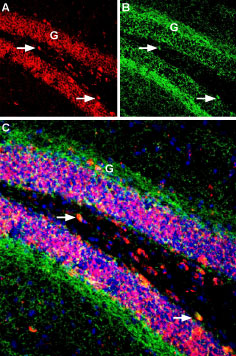
 Multiplex staining of proNGF and Cannabinoid Receptor 1 in mouse hippocampal dentate gyrusImmunohistochemical staining of immersion-fixed, free floating mouse brain frozen sections using Guinea pig Anti-proNGF Antibody (#ANT-005-GP), (1:300) and rabbit Anti-Cannabinoid Receptor 1 (extracellular) Antibody (#ACR-001), (1:300). A. proNGF staining (red) appears in the dentate gyrus (DG) granule layer (G) and in hilar interneurons (arrows). B. CB1R staining (green) appears in axonal processes surrounding the granule layer and a few hilar interneurons (arrows). C. Merge of the two images reveals co-localization of proNGF and CB1R in a few hilar cells. Cell nuclei were stained with DAPI (blue).
