Overview
- Peptide (C)HLRGQRWPFGEAA(S)R, corresponding to amino acid residues 136-150 of human PAR-4 (Accession Q96RI0). Cys 149 was replaced with Ser. 1st extracellular loop.

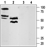
 Western blot analysis of rat lung (lanes 1 and 3) and testis (lanes 2 and 4) lysates:1,2. Anti-PAR4 (F2RL3) (extracellular) Antibody (#APR-034), (1:200).
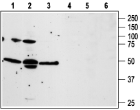
3,4. Anti-PAR4 (F2RL3) (extracellular) Antibody, preincubated with PAR4/F2RL3 (extracellular) Blocking Peptide (#BLP-PR034). Western blot analysis of human prostate carcinoma PC3 (lanes 1 and 4), and Human LNCaP prostate carcinoma (lanes 2 and 5), and human T cell leukemia Jurkat (lanes 3 and 6) cell lines:1-3. Anti-PAR4 (F2RL3) (extracellular) Antibody (#APR-034), (1:200).
4-6. Anti-PAR4 (F2RL3) (extracellular) Antibody, preincubated with PAR4/F2RL3 (extracellular) Blocking Peptide (#BLP-PR034).
 Expression of PAR-4 in rat lungImmunohistochemical staining of rat lung paraffin-embedded sections using Anti-PAR4 (F2RL3) (extracellular) Antibody (APR-034), (1:100). Note that staining is present in the respiratory epithelium of the bronchiole (blue arrow) as well as in the smooth muscle cells of the muscularis mucosae (red arrow). Hematoxilin is used as the counterstain.
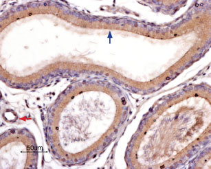
 Expression of PAR-4 in rat epididymisImmunohistochemical staining of rat epididymis paraffin-embedded sections using Anti-PAR4 (F2RL3) (extracellular) Antibody (APR-034), (1:100). Note that strong and specific staining is present in both the pseudostratified epithelium (blue arrow) and the smooth muscle cells of the blood vessels (red arrow). Hematoxilin is used as the counterstain.
 Cell surface detection of PAR-4 by indirect flow cytometry in live intact human MEG-01 megakaryocytic leukemia cells:___ Cells.
___ Cells + goat-anti-rabbit-FITC.
___ Cells + Anti-PAR4 (F2RL3) (extracellular) Antibody (#APR-034), (2.5μg) + goat-anti-rabbit-FITC.- The blocking peptide is not suitable for this application.
 Expression of PAR-4 in a human breast adenocarcinoma cell lineCell surface detection of PAR-4 in a human breast adenocarcinoma cell line. A. Live intact human MCF-7 cells were stained with Anti-PAR4 (F2RL3) (extracellular) Antibody (#APR-034), (1:50), followed by goat anti-rabbit-AlexaFluor-488 secondary antibody (green). B. Live view of the cells. C. Nuclei were visualized with the cell-permeable dye Hoechst 33342 (blue).
- MacFarlane, S.R. et al. (2001) Pharmacol. Rev. 53, 245.
- Hollenberg, M.D. et al. (2002) Pharmacol. Rev. 54, 203.
- Ossovskaya, V.S. et al. (2004) Physiol. Rev. 84, 579.
Protease-activated receptor 4 (PAR-4) belongs to a family of four G protein-coupled receptors (PAR1-4) that are activated as a result of proteolytic cleavage by certain serine proteases, hence their name. In this novel modality of activation, a specific protease cleaves the PAR receptor within a defined sequence in its extracellular N-terminal domain. This results in the creation of a new N-terminal tethered ligand, which subsequently binds to a site in the second extracellular loop of the same receptor. This binding results in the coupling of the receptor to G proteins and in the activation of several signal transduction pathways.1-3
Different PARs are activated by different proteases. Hence, PAR-4 is activated by both thrombin and trypsin whereas PAR-1 and PAR-3 are activated only by thrombin and PAR-2 is activated only by trypsin.1-3 PAR-4 can be also cleaved and activated by other proteases such as cathepsin G.
The intracellular signaling mechanisms mediated by PAR-4 activation are not completely elucidated but they involve calcium mobilization downstream of phospholipase Cβ through the Gαq pathway.1-3
Tissue distribution of PAR-4 is very broad with the highest expression levels found in lung, testis, pancreas and small intestine. In addition, PAR-4 expression was observed in platelets, megakaryocytes and leukocytes. Studies with platelets derived form PAR-4 knockout mice have established an essential role for PAR-4 in thrombin-induced platelet activation. PAR-4 is likely involved in other physiological functions such as regulation of gastrointestinal motility and regulation of vascular endothelial cell function.1-3
Application key:
Species reactivity key:
Anti-PAR4 (F2RL3) (extracellular) Antibody (#APR-034) is a highly specific antibody directed against an extracellular epitope of human protease-activated receptor-4. The antibody can be used in western blot analysis, immunohistochemical, immunocytochemical and flow cytometry applications. It has been designed to recognized PAR-4 from human, mouse and rat samples.
Applications
Citations
- Direct flow cytometry analysis of mouse platelets using #APR-034-F. Tested in PAR4-/- mice platelets.
Arachiche, A. et al. (2013) PLoS ONE 8, e55740.
- Rat dissociated DRGs (1:200).
Chen, D. et al. (2013) Cell. Mol. Neurobiol. 33, 337.
- Sanchez-Centellas, D. et al. (2017) Thromb. Res. 154, 84.
