Overview
- GST fusion protein with the sequence VEDDYTRPPEPVYSTVNKLCDKPASPRHYSPVECDKSFLLSTPY, corresponding to amino acid residues 336-379 of rat Chapsyn-110 (Accession Q63622). Between PDZ2 and PDZ3 domains.

 Western blot analysis of rat brain membranes:1. Anti-PSD-93 Antibody (#APZ-002), (1:1000).
2. Anti-PSD-93 Antibody, preincubated with PSD-93 Blocking Peptide (#BLP-PZ002).
- COS7 cells and mouse brain lysate (Nada, S. et al. (2003) J. Biol. Chem. 278, 47610.).
- Rat brain sections.
- Chick ciliary ganglion cells (Temburni, M.K. et al. (2004) J. Neurosci. 24, 6776.).
- Harris, B.Z. and Lim, W.A. (2001) J. Cell Sci. 114, 3219.
- Hung, A.Y. and Sheng, M. (2002) J. Biol. Chem. 277, 5699.
Chapsyn 110 (also known as PSD-93 and DLG2) is a PDZ containing domain protein that is also a member of the membrane-associated guanylate kinase (MAGUK) family of multi-domain adaptor proteins1,2.
PDZ domains are conserved protein domains of about 90 amino acids involved in protein–protein recognition, protein targeting and assembly of multi-protein complexes. The name PDZ derives from the first three proteins in which these domains were identified: PSD-95 (a 95 kDa protein involved in signaling at the post-synaptic density), DLG (the Drosophila melanogaster Discs Large protein) and ZO-1 (the zonula occludens 1 protein involved in maintenance of epithelial polarity)1,2.
MAGUKs are scaffolding proteins that comprise several modular protein binding motifs including one or more PDZ domains, a Src homology 3 (SH3) domain, and a catalytically inactive guanylate kinase-like domain1,2.
The multidomain nature of PDZ-containing proteins enables them to interact with multiple binding partners and hence organize larger signaling protein complexes.
Indeed, Chapsyn 110 has been shown to participate in the postsynaptic density, a dedicated structure formed in postsynaptic nerve terminals that includes a specialized assembly of ion channels, receptors and signaling molecules that are involved in information processing and the modulation of synaptic plasticity1,2.
Application key:
Species reactivity key:
Anti-PSD-93 Antibody (#APZ-002) is a highly specific antibody directed against an epitope of the rat protein. The antibody can be used in western blot, immunoprecipitation, immunohistochemistry, and immunocytochemistry applications. It has been designed to recognize PSD-93 from rat, mouse, and human samples.

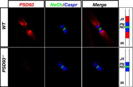
Expression of PSD-93 in mouse sciatic nerve.Immunohistochemical staining of mouse sciatic nerve using Anti-PSD-93 Antibody (#APZ-002). PSD-93 staining (red) is detected in the juxtaparanode region of the Nodes of Ranvier. Shown are Na+ channel staining (green) and Caspr staining (blue) at the node and paranodes. Note the lack of staining in PSD93-/- mice.Adapted from Horresh, I. et al (2008) J. Neurosci. 28, 14213. with permission of the Society for Neuroscience.
Applications
Citations
- Immunohistochemical staining of mouse sciatic nerve. Also tested in PSD93-/- mice.
Horresh, I. et al (2008) J. Neurosci. 28, 14213.
- HEK 293 transfected cells.
Shukla, A. et al. (2017) EMBO J. 36, 458. - Mouse brain lysate.
Lindquist, S. et al (2011) PLoS ONE 6, e23978. - Mouse cortical neuron lysate (1:1000).
Zhang, M. et al (2010) Neuroscience 166, 1083. - Mouse brain lysate (1:1000).
Liaw, W-J. et al (2008) Mol. Pain 4, 45. - Mouse brain lysate.
Jarabek, B.R. et al. (2004) Brain 127, 505. - Chick brain lysate.
Temburni, M.K. et al. (2004) J. Neurosci. 24, 6776. - COS7 cells and mouse brain lysate.
Nada, S. et al. (2003) J. Biol. Chem. 278, 47610.
- COS7 cells and mouse brain lysate.
Nada, S. et al. (2003) J. Biol. Chem. 278, 47610.
- Mouse sciatic nerve. Also tested in PSD93-/- mice.
Horresh, I. et al (2008) J. Neurosci. 28, 14213.
- Chick ciliary ganglion cells.
Temburni, M.K. et al. (2004) J. Neurosci. 24, 6776.
