Overview
- GST fusion protein with the sequence VEDDYTRPPEPVYSTVNKLCDKPASPRHYSPVECDKSFLLSTPY, corresponding to amino acid residues 336-379 of rat Chapsyn-110 (Accession Q63622). Between PDZ2 and PDZ3 domains.
- Rat brain membranes (1:1000-1:2500).
 Western blot analysis of rat brain membranes:1. Anti-PSD-93 Antibody (#APZ-002), (1:1000).
2. Anti-PSD-93 Antibody, preincubated with PSD-93 Blocking Peptide (#BLP-PZ002).
- COS7 cells and mouse brain lysate (Nada, S. et al. (2003) J. Biol. Chem. 278, 47610.).
- Rat brain sections.
- Chick ciliary ganglion cells (Temburni, M.K. et al. (2004) J. Neurosci. 24, 6776.).
Chapsyn 110 (also known as PSD-93 and DLG2) is a PDZ containing domain protein that is also a member of the membrane-associated guanylate kinase (MAGUK) family of multi-domain adaptor proteins1,2.
PDZ domains are conserved protein domains of about 90 amino acids involved in protein–protein recognition, protein targeting and assembly of multi-protein complexes. The name PDZ derives from the first three proteins in which these domains were identified: PSD-95 (a 95 kDa protein involved in signaling at the post-synaptic density), DLG (the Drosophila melanogaster Discs Large protein) and ZO-1 (the zonula occludens 1 protein involved in maintenance of epithelial polarity)1,2.
MAGUKs are scaffolding proteins that comprise several modular protein binding motifs including one or more PDZ domains, a Src homology 3 (SH3) domain, and a catalytically inactive guanylate kinase-like domain1,2.
The multidomain nature of PDZ-containing proteins enables them to interact with multiple binding partners and hence organize larger signaling protein complexes.
Indeed, Chapsyn 110 has been shown to participate in the postsynaptic density, a dedicated structure formed in postsynaptic nerve terminals that includes a specialized assembly of ion channels, receptors and signaling molecules that are involved in information processing and the modulation of synaptic plasticity1,2.
Application key:
Species reactivity key:

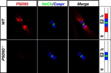
Expression of PSD-93 in mouse sciatic nerve.Immunohistochemical staining of mouse sciatic nerve using Anti-PSD-93 Antibody (#APZ-002). PSD-93 staining (red) is detected in the juxtaparanode region of the Nodes of Ranvier. Shown are Na+ channel staining (green) and Caspr staining (blue) at the node and paranodes. Note the lack of staining in PSD93-/- mice.Adapted from Horresh, I. et al (2008) J. Neurosci. 28, 14213. with permission of the Society for Neuroscience.
