Overview
- Peptide RNGTRGGNDTFKC, corresponding to amino acids 161-173 of rat HRH2 (Accession P25102). 2nd extracellular loop.

 Western blot analysis of rat basophilic leukemia cells (RBL) (lanes 1 and 3) and rat brain lysates (lanes 2 and 4):1,2. Anti-Histamine H2 Receptor (HRH2) (extracellular) Antibody (#AHR-002), (1:200).
3,4. Anti-Histamine H2 Receptor (HRH2) (extracellular) Antibody, preincubated with Histamine H2 Receptor/HRH2 (extracellular) Blocking Peptide (#BLP-HR002).
 Expression of Histamine H2 receptor in rat stomachImmunohistochemical staining of paraffin embedded section of rat stomach using Anti-Histamine H2 Receptor (HRH2) (extracellular) Antibody (#AHR-002), (1:100). H2R (brown staining) is expressed in parietal cells (arrows) of the gastric glands. Hematoxilin is used as the counterstain.
- Mouse primary microglia (Apolloni, S. et al. (2017) Front. Immunol. 8, 1689.).
 Cell surface detection of Histamine H2 receptor in live intact rat basophilic leukemia cell lines:___ Unstained cells + goat-anti-rabbit-FITC.
___ Cells+ Anti-Histamine H2 Receptor (HRH2) (extracellular) Antibody (#AHR-002), (1:20) + goat-anti-rabbit-FITC.- The control antigen is not suitable for this application.
- Hill, S.J. et al. (1997) Pharmacol. Rev. 49, 253.
- Del Valle, J. and Gantz, I. (1997) Am. J. Physiol. 273, G987.
- Kobayashi, T. et al. (2000) J. Clin. Invest. 105, 1741.
Histamine (2-[4-imidazole]ethylamine) is a low-molecular-weight amine synthesized from L-histidine. It is produced by various cells throughout the body, including central nervous system neurons, gastric mucosa parietal cells, mast cells, basophils and lymphocytes. Histamine is a major biological mediator whose functions include, among many others, regulation of vascular smooth muscle, immune regulation, regulation of sleep-wake cycles and regulation of gastric acid secretion.1,2
The biological effects of histamine are mediated through four receptors (H1-H4 receptors) all of which belong to the 7-transmembrane domain, G protein-coupled receptor (GPCR) superfamily.
H2 receptors couple to adenylate cyclase via Gs, leading to the formation of cAMP and the activation of protein kinase A (PKA).1,2 H2 receptors are expressed in several tissues including the brain, gastrointestinal tract and heart.
The most studied physiological role of the H2 receptor is its involvement in the regulation of gastric acid secretion.3 Specific H2 receptor antagonists are currently used for the treatment of acid-related gastrointestinal conditions such as peptic ulcer, gastroesophageal reflux disease and dyspepsia.
Involvement of H2 receptors in chronic heart failure and immune regulation has also been suggested.
Application key:
Species reactivity key:
Alomone Labs is pleased to offer a highly specific antibody directed against an epitope of rat histamine H2 receptor. Anti-Histamine H2 Receptor (HRH2) (extracellular) Antibody (#AHR-002) can be used in western blot, indirect flow cytometry, immunohistochemistry, and immunocytochemistry applications. The antibody recognizes an extracellular epitope and is thus ideal for detecting the receptor in living cells. It has been designed to recognize H2R from rat and mouse samples. It will not recognize human H2R.

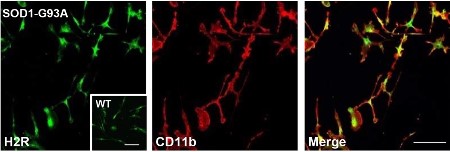
Expression of Histamine H2 Receptor in mouse primary microglia.Immunocytochemical staining of mouse primary microglia using Anti-Histamine H2 Receptor (HRH2) (extracellular) Antibody (#AHR-002). H2R expression (green) partially co-localizes with CD11b (red).Adapted from Apolloni, S. et al. (2017) Front. Immunol. 8, 1689. with permission of Frontiers.
Applications
Citations
- Rat primary astrocyte lysate (1:200).
Xu, J. et al. (2018) J. Neuroinflammation 15, 41. - Mouse brain and spinal cord lysate.
Apolloni, S. et al. (2017) Front. Immunol. 8, 1689.
- Mouse intestine sections.
Blasco, M.P. et al. (2020) J. Neurounflammation 17, 160. - Mouse intestine sections.
Gao, C. et al. (2015) mBio 6, e01358.
- Rat primary astrocyte lysate (1:200).
Xu, J. et al. (2018) J. Neuroinflammation 15, 41. - Mouse primary microglia.
Apolloni, S. et al. (2017) Front. Immunol. 8, 1689.
- Mouse P815 mast cells.
Dong, H. et al. (2019) Front. Cell. Neurosci. 13, 191.
