Overview
- Solubilized proteins from PC12 membranes.
P07174
24596
- PC12 lysate (non-reduced) 1:500-1:2500.
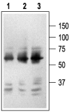
 Western blot analysis of PC12 cell lysate (non-reduced):1. Mouse Anti-Rat p75 NGF Receptor (extracellular) Antibody (#AN-170), (1:2500).
2. Mouse Anti-Rat p75 NGF Receptor (extracellular) Antibody (1:1500).
3. Mouse Anti-Rat p75 NGF Receptor (extracellular) Antibody (1:150).
- Rat brain sections (1:300).
- PC12 cells.
p75NTR known as a cell surface receptor for NGF, BDNF, NT-3 and NT-4, is also a receptor for b-amyloid, and for the myelin axon repellant protein MAG.1,2,4,6 Binding of neurotrophins induces receptor dimerization followed by phosphorylation of receptor kinase residues and recruitment of intracellular proteins involved in signal transduction.
p75NTR functions alone, or in functional complexes with the neurotrophin receptors trkA, trkB and trkC. p75NTR signals via interaction with different transducers such as TRAF6, IRAK, and RIP-2, activating NF-κB, JNK and p38, and inactivating rhoA.1,3,6
Receptor degradation is mediated by a metalloproteinase-dependent shedding of the extracellular domain. p75NTR binds all neurotrophins with similar nM affinity, but binds pro-neurotrophins with greater affinity, and is more potently activated by pro-neurotrophins.5
Overexpression of the intracellular domain in developing neurons induces apoptosis. NGF-dependent association of NRIF with p75NTR contributes to pro-apoptotic signaling.7,8
Beta-amyloid peptide binds p75NTR and stimulates pro apoptotic signaling by p75NTR, suggesting that the receptor may be relevant for Alzheimer's disease.9
Application key:
Species reactivity key:
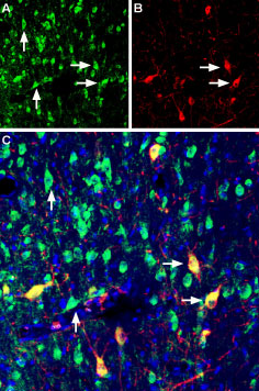
 Multiplex staining of Vesicular Acetylcholine Transporter and p75NTR in rat medial septum.Immunohistochemical staining of immersion-fixed, free floating rat brain frozen sections using rabbit Anti-Vesicular Acetylcholine Transporter (VAChT) Antibody (#ACT-003), (1:200) and Mouse Anti-Rat p75 NGF Receptor (extracellular) Antibody (#AN-170), (1:200). A. VAChT staining (green) appears in several neuronal cells. B. p75NTR (red) also stains neuronal cells. C. Merge of the two images shows colocalization of VAChT and p75NTR in some cells (horizontal arrows), while other cells express only VAChT (vertical arrows). Cell nuclei are stained with DAPI (blue).
