Overview
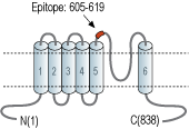
- Peptide (C)NSLPMESTPHK*SRGS, corresponding to amino acid residues 605-619 of rat TRPV1 with replacement of cysteine 616 (C616) with serine (*S) (Accession O35433). 3rd extracellular loop.

Won't recognize TRPV1 from human samples.
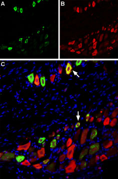
 Multiplex staining of TRPV1 and NaV1.8 in rat DRGImmunohistochemical staining of rat dorsal root ganglion (DRG) using Anti-TRPV1 (VR1) (extracellular)-ATTO Fluor-488 Antibody (#ACC-029-AG), (green), (1:60) and Anti-NaV1.8 (SCN10A)-ATTO Fluor-594 Antibody (#ASC-016-AR), (red), (1:60). A. TRPV1 staining. B. NaV1.8 staining. C. Merge of A and B demonstrates partial co-localization of TRPV1 and NaV1.8 channels. Nuclei stained using DAPI as the counterstain (blue).
 Expression of TRPV1 in rat DRGImmunohistochemical staining of rat dorsal root ganglion (DRG) frozen sections using Anti-TRPV1 (VR1) (extracellular )-ATTO Fluor-488 Antibody (#ACC-029-AG), (1:50), (green). TRPV1 is expressed in medium and small DRG neurons. Hoechst 33342 is used as the counterstain (blue).
 Expression of TRPV1 in rat PC12 cellsCell surface detection of TRPV1 in intact living Pheochromocytoma (PC12) cells. A. Extracellular staining of cells using Anti-TRPV1 (VR1) (extracellular)-ATTO Fluor-488 Antibody (#ACC-029-AG), (1:25), (green). B. Live view of the cell. C. Merge images of A and B.
- Montell, C. et al. (2002) Mol. Cell. 9, 229.
- Clapham, D.E. (2003) Nature 426, 517.
- Moran, M.M. et al. (2004) Curr. Opin. Neurobiol. 14, 362.
- Clapham, D.E. et al. (2003) Pharmacol. Rev. 55, 591.
- Gunthorpe, M.J et al. (2002) Trends. Pharmacol. Sci. 23, 183.
- Ross, R.A. et al. (2003) Br. J. Pharmacol. 140, 790.
- Tominaga, M. et al. (1998) Neuron 21, 531.
- Agopyan, N. et al. (2004) Am. J. Physiol. 286, L563.
- Reilly, C.A. et al. (2003) Toxicol. Sci. 73, 170.
TRP channels are a large family (about 28 genes) of plasma membrane, non-selective cationic channels that are either specifically or ubiquitously expressed in excitable and non-excitable cells.1
According to IUPHAR the TRP family comprises three main subfamilies on the basis of sequence homology; TRPC, TRPM and TRPV (to date, three extra subfamilies are considered to belong to the TRP family; the TRPA, TRPML, and TRPP).1-4 The TRPV subfamily consists of six members, TRPV1-6.5
TRPV1 channel has many activators; among them heat, protons, vanilloids like capsaicin, resiniferatoxin (RTX), and lipids. This channel is associated with tissue injury and inflammation.6,7 TRPV1 is expressed predominantly in nociceptors and in sensory neurons.
Recent studies demonstrated involvement of TRPV1 in apoptosis where inhibition of the receptor prevented apoptosis.8,9
Application key:
Species reactivity key:
Anti-TRPV1 (VR1) (extracellular) Antibody (#ACC-029) is a highly specific antibody directed against an extracellular epitope of the rat TRPV1 channel. The antibody can be used in western blot, immunohistochemistry, and live cell imaging applications. It has been designed to recognize TRPV1 from rat and mouse samples. The antibody will not work with human samples.
Anti-TRPV1 (VR1) (extracellular)-ATTO Fluor-488 Antibody (#ACC-029-AG) is directly labeled with an ATTO-488 fluorescent dye. ATTO dyes are characterized by strong absorption (high extinction coefficient), high fluorescence quantum yield, and high photo-stability. The ATTO-488 label is analogous to the well known dye fluorescein isothiocyanate (FITC) and can be used with filters typically used to detect FITC. Anti-TRPV1 (VR1) (extracellular)-ATTO Fluor-488 Antibody has been tested in immunohistochemistry and live cell imaging applications and is especially suited for experiments requiring simultaneous labeling of different markers.
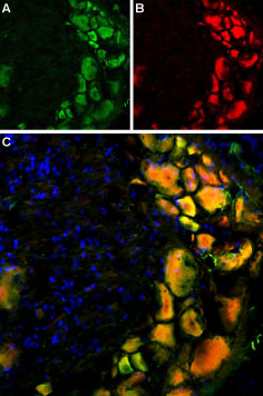
 Multiplex staining of TRPV1 and mGluR5 in rat DRGImmunohistochemical staining of rat dorsal root ganglion using Anti-TRPV1 (VR1) (extracellular)-ATTO Fluor-488 Antibody (#ACC-029-AG), (1:60) and Anti-mGluR5 (extracellular)-ATTO Fluor-594 Antibody (#AGC-007-AR), (1:60). A. TRPV1 staining (green). B. mGluR5 staining of the same section (red). C. Merge of A and B demonstrates co-localization of TRPV1 and mGluR5 in DRG cells. Nuclei were stained using DAPI as the counterstain (blue).

