Overview
- Peptide (C)RAENEKDATTEKNKKR, corresponding to amino acid residues 1371-1386 of human RyR1 (Accession P21817). Intracellular, N-terminus.

 Western blot analysis of rat skeletal muscle lysates:1. Anti-Ryanodine Receptor 1 Antibody (#ARR-001), (1:200).
2. Anti-Ryanodine Receptor 1 Antibody, preincubated with Ryanodine Receptor 1 Blocking Peptide (#BLP-RR001).
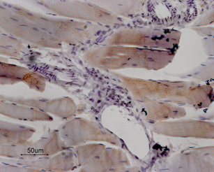
 Expression of RyR1 in rat skeletal muscleImmunohistochemical staining of paraffin embedded sections of rat quadriceps using Anti-Ryanodine Receptor 1 Antibody (#ARR-001), (1:100). RyR1 is expressed in fibers of striate muscle. Note that smooth muscle in the arterial walls is negative as well as the surrounding connective tissue. Hematoxilin is used as the counterstain.
 Expression of RyR1 in mouse cerebellumImmunohistochemical staining of mouse cerebellum frozen sections using Anti-Ryanodine Receptor 1 Antibody (#ARR-001), (1:100), (green). A. The highest expression of RyR1 is in the molecular layer (asterisk). B. In the same section, there is staining for parvalbumin (red), a marker of Purkinje cells. C. Merged image of panels A and B demonstrates that RyR1 is localized surrounding the dendritic tree of Purkinje cells. DAPI is used as the counterstain.
- HEK-293 cells transfected with rabbit RyR1 (1:500) (Kaya, L. et al. (2013) Biochim. Piophys. Acta 1833, 1421.)
- Chakrabarti, R. and Chakrabarti, R. (2006) J. Cell. Biochem. 99, 1503.
- Eisner, D.A. et al. (2005) Exp. Physiol. 90, 3.
- Hamilton, S.L. (2005) Cell Calcium 38, 253.
- Fill, M. and Copello, J.A. (2002) Physiol. Rev. 82, 893.
It is well established that cytosolic calcium (Ca2+) acts as a key second messenger in many intracellular pathways including synaptic transmission, muscle contraction, hormonal secretion, cell growth and proliferation.1,2 The primary intracellular Ca2+ storage/release organelle in most cells is the endoplasmic reticulum (ER) or the sarcoplasmic reticulum (SR) in striated muscle cells.
The ER and SR contain two Ca2+ release channel families, the inositol trisphosphate receptors (IP3Rs) and the ryanodine receptors (RyRs).3
The ryanodine receptor family consists of three different isoforms: the skeletal muscle isoform, ryanodine receptor type 1 (RyR1); the cardiac muscle isoform, ryanodine receptor type 2 (RyR2) and the brain isoform, ryanodine receptor type 3 (RyR3).3
The ryanodine receptors are homotetrameric proteins, which upon assembly create a Ca2+ conducting channel. They play a key role in the mechanism of excitation-contraction coupling in striated muscle. Binding of ryanodine (a poisonous alkaloid found in the South American plant Ryania speciosa) to the ryanodine receptor causes two major changes in the channel: a reduction in single-channel conductance and a marked increase in the open state probability, leading to an overall increase/decrease in the Ca2+ release capability of the channel.
RyR1 is expressed predominantly in skeletal muscle and in areas of the brain; human RyR1 has at least three known alternative splice variants.
Several diseases are attributed to mutations in the RyR1 gene: Central core disease (CCD), multi-minicore disease (MmD) and Malignant hyperthermia (MH) 4.
Application key:
Species reactivity key:
Anti-Ryanodine Receptor 1 Antibody (#ARR-001) is a highly specific antibody directed against an epitope of human RyR1. The antibody can be used in western blot, immunocytochemistry, and immunohistochemistry applications. It has been designed to recognize RyR1 from human, rat, and mouse samples.

Expression of RyR1 in rat tibialis anterior muscle.Immunohistochemical staining of rat tibialis anterior muscle sections using Anti-Ryanodine Receptor 1 Antibody (#ARR-001). RyR1 co-localizes with Calpain II.Adapted from Kraner, S.D. et al. (2011) Am. J. Physiol. 300, R1384. with permission of The American Physiological Society.
Applications
Citations
- Rat brain lysate (1:1000).
Kaya, L. et al. (2013) Biochim. Piophys. Acta 1833, 1421. - Rat cardiac myoblast cell lysate (H9c2).
O-Uchi, J. et al. (2013) Am. J. Physiol. 305, H1736. - Rat intrapulmonary artery lysate (1:200).
Dahan, D. et al. (2012) Am. J. Physiol. 303, L824. - Rat muscle lysate.
Kraner, S.D. et al. (2011) Am. J. Physiol. 300, R1384.
- Rat tibialis anterior muscle sections (1:100).
Kraner, S.D. et al. (2011) Am. J. Physiol. 300, R1384.
- Rat adipose-derived stromal cells and rat bone marrow stromal cells.
Forostyak, O. et al. (2016) Stem Cell Res. 16, 622. - Human ESC NPs (1:200).
Forostyak, O. et al. (2013) Stem Cells Dev. 22, 1506. - HEK-293 cells transfected with rabbit RyR1 (1:500).
Kaya, L. et al. (2013) Biochim. Piophys. Acta 1833, 1421.
- Rebello, M.R. and Medler, K.F. (2010) Eur. J. Neurosci. 32, 1825.
- Yoshida, S. et al. (2010) Neurosci. Res. 68, 322.
