Overview
- Peptide (C)DYVGKLAGRLRD, corresponding to amino acid residues 23-34 of mouse S1PR3 (Accession Q9Z0U9). Extracellular, N-terminus.
- Mouse J774 macrophage and human Jurkat T-cell leukemia cells (2.5-5 µg).
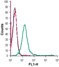
 Cell surface detection of S1PR3 in live intact mouse J774 macrophage cells:___ Cells.
___ Cells + rabbit IgG isotype control-FITC.
___ Cells + Anti-S1PR3 (EDG3) (extracellular)-FITC Antibody (#ASR-013-F), (2.5 µg). Cell surface detection of S1PR3 in live intact human Jurkat T-cell leukemia cells:___ Cells.
___ Cells + rabbit IgG isotype control-FITC.
___ Cells + Anti-S1PR3 (EDG3) (extracellular)-FITC Antibody (#ASR-013-F), (5 µg).
Sphingosine 1-phosphate (S1P) is an active byproduct of sphingomyelin metabolism. All cells can synthesize this biomolecule but the majority of its synthesis comes from erythrocytes and endothelial cells. In cases of inflammation, mast cells and platelets are the main source of sphingosine 1-phosphate1. S1P cellular functions could be intracellular, where it is synthesized. In addition, upon secretion, it circulates in the blood via its binding to high-density lipoproteins and albumin. When S1P reaches its target cell it could activate 5 different high affinity receptors belonging to the G-protein coupled receptor superfamily: Sphingosine 1-phosphate receptors, termed S1PR1-51.
Stimulation of S1P receptors triggers a cascade of signaling events depending on the receptor and on the G-protein it couples. S1PR1 couples to Gi2. S1PR2 can couple to Gs, Gq or G12/13 and S1PR3-5 can couple to Gior G12/133-4. The pathways activated vary from Ca2+ mobilization, activation or inhibition of adenylate cyclase phospholipase C activation and more. Through the different signaling pathways these receptors activate, S1P1 receptors are implicated in adherens junction assembly, cytoskeletal changes, cell migration, proliferation and apoptosis5.
S1PR3 is specifically detected in the brain, heart, spleen, liver, lung, thymus, kidney, testis and skeletal muscle3. S1PR1 and S1PR2 are generally expressed in the CNS, cardiovascular and immune systems1. S1PR4 is specifically expressed in the lymphoid tissue and S1PR5 in natural killer cells and olygodendrocytes1.
Malfunction of S1P receptor signaling is reported in various disorders, for example multiple sclerosis and may be targets for the development of therapeutic drugs1.
