Overview
- Peptide (C)NHQDKKGVIRESYLK, corresponding to amino acid residues 626 - 640 of mouse Semaphorin 6A (Accession O35464). Extracellular, N-term.
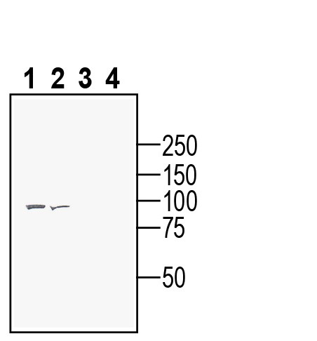
 Western blot analysis of rat brain membranes (lanes 1 and 3) and mouse brain membranes (lanes 2 and 4):1-2. Anti-Semaphorin 6A (extracellular) Antibody (#ASR-071), (1:200).
3-4. Anti-Semaphorin 6A (extracellular) Antibody, preincubated with Semaphorin 6A (extracellular) Blocking Peptide (BLP-SR071). Western blot analysis of human Malme-3M melanoma cell line lysate (lanes 1 and 3) and human Colo-205 colorectal adenocarcinoma cell line lysate (lanes 2 and 4):1-2. Anti-Semaphorin 6A (extracellular) Antibody (#ASR-071), (1:200).
3-4. Anti-Semaphorin 6A (extracellular) Antibody, preincubated with Semaphorin 6A (extracellular) Blocking Peptide (BLP-SR071).
Semaphorin 6A (Sema6A) is a member of the semaphorin family of genes involved in cell migration, axon guidance, development of bone1,2, vasculature3, cancer metastasis4, B-cell aggregation and differentiation5, and synaptogenesis6-12, as well as in inhibiting synaptic terminal arborization13.
The semaphorins represent one of the largest families of axon guidance cues consisting of eight classes. Members are classified according to structural criteria. All semaphorins share a highly conserved Sema-domain of about 500 amino acids14 with 17 conserved cysteines. Classes 1 and 2 are found only in invertebrates, while classes 3 to 7 contain the vertebrate family members, and class V members are encoded by viral genomes. Semaphorins include both transmembrane (classes 1, 4, 5 and 6), glycosylphosphatidyl inositol (class 7) and secreted (classes 2, 3 and V) proteins15.
Initially, all semaphorin family members were believed to induce inhibitory actions on axon pathfinding, branching or targeting, but there is increasing evidence that semaphorins may also have a role in chemoattraction13,16-19. Mutations of Sema6A in mice is associated with a spectrum of subtle defects in cell migration and axon guidance in various brain areas, including the thalamocortical system, hippocampus, cerebellum and various other structures6-12,20. Previous study of gene expression analysis revealed alterations in semaphorin and plexin expression in the prefrontal cortex of patients with schizophrenia20.
